Review
. 2016 Oct;33(10):1279-1286.
doi: 10.1007/s10815-016-0768-3. Epub 2016 Jul 16.

Implementing PGD/PGD-A in IVF clinics: considerations for the best laboratory approach and management

Affiliations
Review

Implementing PGD/PGD-A in IVF clinics: considerations for the best laboratory approach and management

Antonio Capalbo et al. J Assist Reprod Genet. 2016 Oct.

Abstract

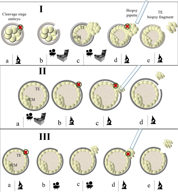
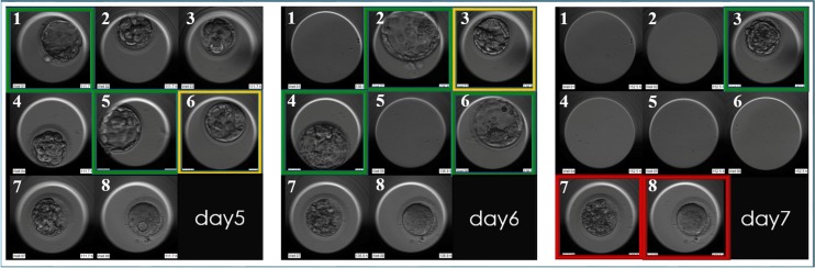
For an IVF clinic that wishes to implement preimplantation genetic diagnosis for monogenic diseases (PGD) and for aneuploidy testing (PGD-A), a global improvement is required through all the steps of an IVF treatment and patient care. At present, CCS (Comprehensive Chromosome Screening)-based trophectoderm (TE) biopsy has been demonstrated as a safe, accurate and reproducible approach to conduct PGD-A and possibly also PGD from the same biopsy. Key challenges in PGD/PGD-A implementation cover genetic and reproductive counselling, selection of the most efficient approach for blastocyst biopsy as well as of the best performing molecular technique to conduct CCS and monogenic disease analysis. Three different approaches for TE biopsy can be compared. However, among them, the application of TE biopsy approaches, entailing the zona opening when the expanded blastocyst stage is reached, represent the only biopsy methods suited with a totally undisturbed embryo culture strategy (time lapse-based incubation in a single media). Moreover, contemporary CCS technologies show a different spectrum of capabilities and limits that potentially impact the clinical outcomes, the management and the applicability of the PGD-A itself. In general, CCS approaches that avoid the use of whole genome amplification (WGA) can provide higher reliability of results with lower costs and turnaround time of analysis. The future perspectives are focused on the scrupulous and rigorous clinical validations of novel CCS methods based on targeted approaches that avoid the use of WGA, such as targeted next-generation sequencing technology, to further improve the throughput of analysis and the overall cost-effectiveness of PGD/PGD-A.

Keywords: CCS; Embryo selection; PGD; PGD-A; PGS; Trophectoderm biopsy.


Figures

Fig. 1
Fig. 1
Blastocyst biopsy methods. I. Day3 hatching-based blastocyst biopsy method. (a) The embryo is taken out from the incubator on day3 of preimplantation development and the zona pellucida is drilled; (b) The embryo is culture up to the blastocyst stage either in time-lapse or in a standard incubator; (c) The trophectoderm cells (or in some cases also the Inner Cell Mass) will herniate from the opening in the zona pellucida; (d) The biopsy fragment is easily collected by firing with the laser at the junctions between the cells at the edges and the body of the blastocyst; (e) The biopsy fragment is released. II. Sequential zona opening and blastocyst biopsy method. (a) The embryo is cultured up to the fully-expanded blastocyst stage either in time-lapse or in a standard incubator; (bc) The zona pellucida is drilled in a spot opposite to the Inner Cells Mass, the trophectoderm is gently detached from the it and few cells are sucked within the pipette to be then removed from the body of the blastocyst by alternating stretching and firing the laser; (d) The biopsy fragment is released. III. Day5/6 hatching-based blastocyst biopsy method. (a) The blastocyst is taken out from the incubator in order to drill the zona pellucida on day5 or day6 of preimplantation development. Importantly, the inner cell mass is kept opposite to the spot where zona is opened; (bc) The blastocyst is put back into the incubator until few trophectoderm cells will herniate; (d) The biopsy fragment is easily collected by firing with the laser at the junctions between the cells at the edges and the body of the blastocyst; (e) The biopsy fragment is released. The microscope icon identifies the steps conducted under the laser-equipped micromanipulator; the camera icon identifies the steps preferably conducted in a time-lapse system; the steps identified by the incubator and camera icons can be either conducted in a time-lapse system or in a conventional incubator. ICM, Inner Cell Mass; TE, trophectoderm; green circle with inner red cross, laser’s target
Fig. 2
Fig. 2
The blastocyst biopsy approaches that omit the day 3 hatching step guarantee a better synchronization between physiological embryo development and daily laboratory management. In this example in day 5 of preimplantation development, embryo n1 and n5 reach full expansion and undergo trophectoderm (TE) biopsy and cryopreservation soon after. Embryo n6 is kept in culture in a time-lapse instrument up to day6. In day 6, embryo n2, n4 and indeed n6 undergo TE biopsy and cryopreservation. Embryo n3 is given a last chance to develop as a fully expanded blastocyst, and kept in culture up to day 7, when it is finally biopsied and cryopreserved. Green boxes identify blastocyst selected for biopsy; orange boxes identify blastocysts that are kept in culture in time-lapse on each specific day of preimplantation development; red boxes identify embryos that did not develop as blastocysts

References

    1. Handyside AH, Kontogianni EH, Hardy K, Winston RM. Pregnancies from biopsied human preimplantation embryos sexed by Y-specific DNA amplification. Nature. 1990;344(6268):768–770. doi: 10.1038/344768a0. - DOI - PubMed
    1. Lee E, Illingworth P, Wilton L, Chambers GM. The clinical effectiveness of preimplantation genetic diagnosis for aneuploidy in all 24 chromosomes (PGD-A): systematic review. Hum Reprod. 2015;30(2):473–483. doi: 10.1093/humrep/deu303. - DOI - PubMed
    1. Dahdouh EM, Balayla J, García-Velasco JA. Comprehensive chromosome screening improves embryo selection: a meta-analysis. Fertil Steril. 2015;104(6):1503–1512. doi: 10.1016/j.fertnstert.2015.08.038. - DOI - PubMed
    1. Dahdouh EM, Balayla J, Audibert F, Genetics Committee. Wilson RD, Audibert F, Brock JA, Campagnolo C, Carroll J, Chong K, Gagnon A, Johnson JA, MacDonald W, Okun N, Pastuck M, Vallée-Pouliot K. Technical update: preimplantation genetic diagnosis and screening. J Obstet Gynaecol Can. 2015;37(5):451–63. doi: 10.1016/S1701-2163(15)30261-9. - DOI - PubMed
    1. Chen M, Wei S, Hu J, Quan S. Can comprehensive chromosome screening technology improve IVF/ICSI outcomes? A Meta-Analysis. PLoS One. 2015;10(10) - PMC - PubMed