Review
. 2016 Dec:60:89-96.
doi: 10.1016/j.semcdb.2016.07.015. Epub 2016 Jul 14.

Membrane fusion during poxvirus entry

Affiliations
Review

Membrane fusion during poxvirus entry

Bernard Moss. Semin Cell Dev Biol. 2016 Dec.

Abstract

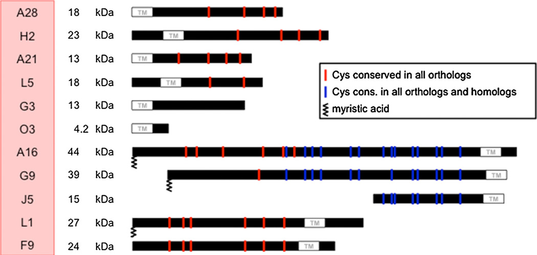
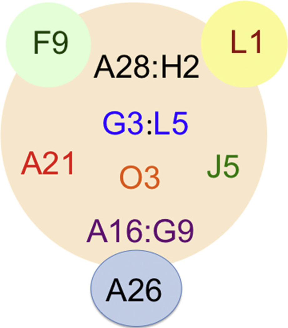
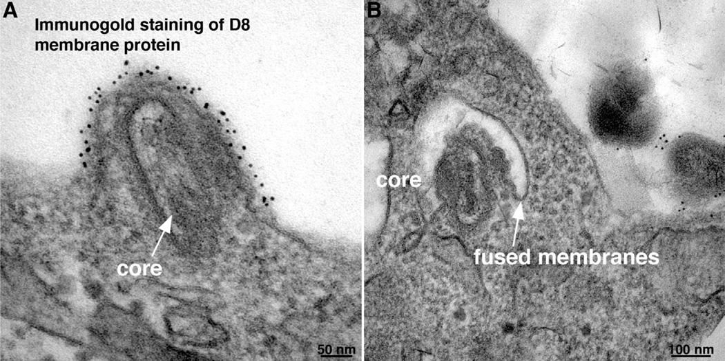
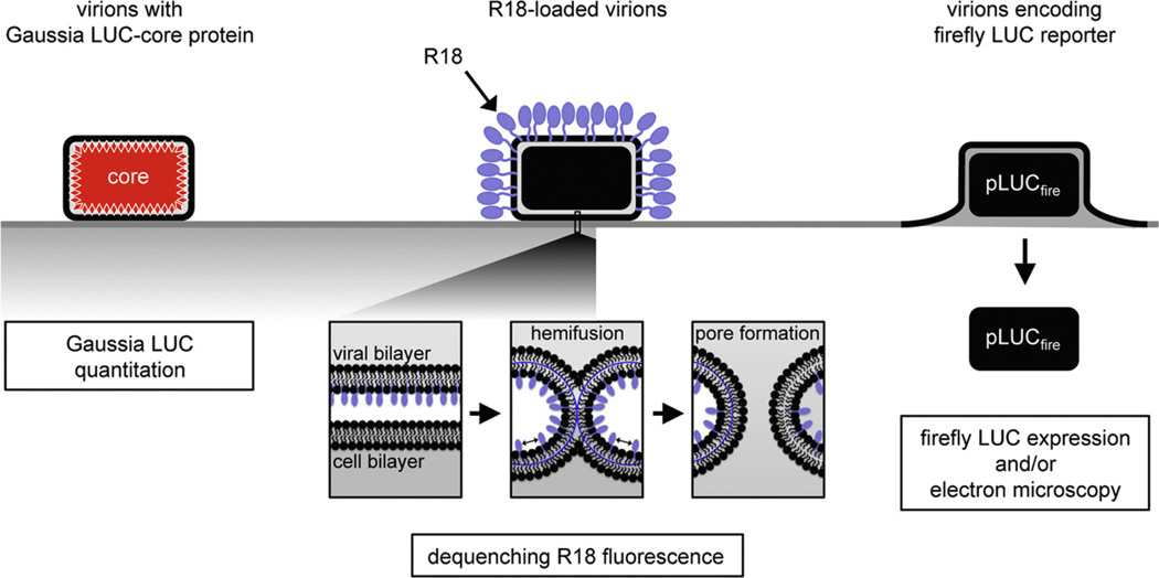
Poxviruses comprise a large family of enveloped DNA viruses that infect vertebrates and invertebrates. Poxviruses, unlike most DNA viruses, replicate in the cytoplasm and encode enzymes and other proteins that enable entry, gene expression, genome replication, virion assembly and resistance to host defenses. Entry of vaccinia virus, the prototype member of the family, can occur at the plasma membrane or following endocytosis. Whereas many viruses encode one or two proteins for attachment and membrane fusion, vaccinia virus encodes four proteins for attachment and eleven more for membrane fusion and core entry. The entry-fusion proteins are conserved in all poxviruses and form a complex, known as the Entry Fusion Complex (EFC), which is embedded in the membrane of the mature virion. An additional membrane that encloses the mature virion and is discarded prior to entry is present on an extracellular form of the virus. The EFC is held together by multiple interactions that depend on nine of the eleven proteins. The entry process can be divided into attachment, hemifusion and core entry. All eleven EFC proteins are required for core entry and at least eight for hemifusion. To mediate fusion the virus particle is activated by low pH, which removes one or more fusion repressors that interact with EFC components. Additional EFC-interacting fusion repressors insert into cell membranes and prevent secondary infection. The absence of detailed structural information, except for two attachment proteins and one EFC protein, is delaying efforts to determine the fusion mechanism.

Keywords: Endocytosis; Hemifusion; Membrane fusion; Vaccinia virus; Virus entry.


Figures

Fig. 1
Fig. 1. Protein components of the EFC
The locations of the transmembrane domains of the 11 proteins comprising the EFC are shown. Nine of the proteins have cysteines that are conserved in all poxirus orthologs. Three proteins (A16, G9 and J5) are homologs derived by duplications. Three proteins (A16, G9 and L1) are myristoylated.
Fig. 2
Fig. 2. Interactions of components of EFC
Of the eleven EFC components, nine (A16, A21, A28, G3, G9, H2, J5, L5 and O3) constitute the core and two (F9 and L1) are peripherally located. The EFC proteins that are known to interact directly (A28:H2, G3:L5, and A16:G9) are shown in the same color separated by a colon. The A26 fusion suppressor protein interacts with the A16:G9 subcomplex.
Fig. 3
Fig. 3. Fusion of vaccinia virus mature virions with cells visualized by transmission electron microscopy
Immunogold staining with antibody to the D8 membrane protein was performed prior to embedding and cryosectioning. (A) Fusion at the plasma membrane 10 min after infection. Arrow points to virus core. (B) Fusion with endosomal membrane 20 min after infection. Arrow points to junction of viral and endosomal membrane. Modified from: Townsley, A.C., et al. (2006). J. Virol. 80, 8899–8908.
Fig. 4
Fig. 4. Measurement of binding, hemifusion and core entry steps
Attachment: a recombinant VACV with Gaussia luciferase (LUC) fused to a core protein is used to measure the binding of virions at 4 °C by assaying cell-associated LUC activity. Hemifusion: R18-loaded virions are bound to target cells at 4 °C, shifted to 37 °C, and the dequenching of R18 dimers due to lipid mixing is measured by increased fluorescence. Core entry: a recombinant VACV that expresses firefly LUC under an early promoter, is used to infect cells and newly synthesized LUC is measured. Modified from: Laliberte, J.P., Weisberg, A.S., Moss, B., 2011. PLoS Pathog. 7, e1002446.

Similar articles

Cited by

References

    1. White JM, Delos SE, Brecher M, Schornberg K. Structures and mechanisms of viral membrane fusion proteins: multiple variations on a common theme. Crit. Rev. Biochem. Mol. Biol. 2008;43(3):189–219. - PMC - PubMed
    1. Chernomordik LV, Kozlov MM. Membrane hemifusion: crossing a chasm in two leaps. Cell. 2005;123(3):375–382. - PubMed
    1. Moss B. Poxvirus cell entry: how many proteins does it take? Viruses. 2012;4(5):688–707. - PMC - PubMed
    1. Laliberte JP, Moss B. Lipid membranes in poxvirus replication. Viruses Basel. 2010;2(4):972–986. - PMC - PubMed
    1. Schmidt FI, Bleck CK, Mercer J. Poxvirus host cell entry. Curr. Opin. Virol. 2012;2(1):20–27. - PubMed

Publication types

LinkOut - more resources