

Isolated hemangioblastoma of the cervical spinal cord: A case report and literature review
- PMID: 27424104
- PMCID: PMC4949809
- DOI: 10.1016/j.ijscr.2016.07.002
Isolated hemangioblastoma of the cervical spinal cord: A case report and literature review
Abstract
Introduction: Hemangioblastomas are benign, slow growing but highly vascularized tumors of the central nervous system, with the most common location of occurrence in the posterior fossa. Hemangioblastomas usually have an associated with patients that have Von-Hippel Lindau disease, resulting a germline mutation in the VHL tumor suppressor gene. Isolated or sporadic occurrences of hemangioblastomas are much more infrequent and typically respond well after surgery.
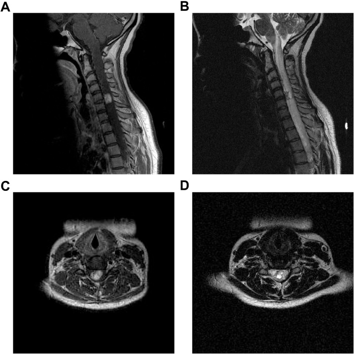
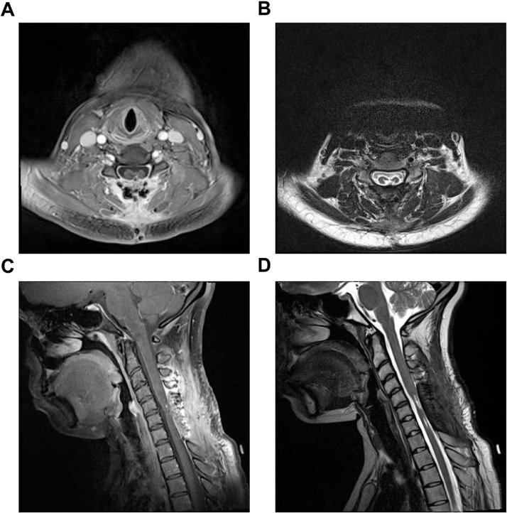
Presentation of case: We present case of a 22year old female with worsening shoulder pain, decreased sensation in the hands and feet, and decreasing strength and was found to have a hemangioblastoma of the cervical spine.
Discussion: The patient was treated with surgery and responded well to treatment. We also present a review of the literature on isolated occurrences of hemangioblastomas of the spinal cord.
Conclusion: Isolated hemangioblastoma are a rare tumor of the central nervous system and can be managed with surgery.
Keywords: Case report; Hemangioblastoma; Laminectomy; Myelotomy; Vascular tumor; Von-Hippel Lindau.
Published by Elsevier Ltd.
Figures
Similar articles
-
Sporadic and Von-Hippel Lindau disease-associated spinal hemangioblastomas: institutional experience on their similarities and differences.J Neurooncol. 2019 Jul;143(3):547-552. doi: 10.1007/s11060-019-03189-w. Epub 2019 May 14. J Neurooncol. 2019. PMID: 31089924
-
Extraneuraxial hemangioblastoma: A clinicopathologic study of 10 cases with molecular analysis of the VHL gene.Pathol Res Pract. 2018 Aug;214(8):1156-1165. doi: 10.1016/j.prp.2018.05.007. Epub 2018 May 29. Pathol Res Pract. 2018. PMID: 29941223
-
Comparison of anterior and posterior surgical approaches in the treatment of ventral spinal hemangioblastomas in patients with von Hippel-Lindau disease.J Neurosurg. 2003 Jan;98(1):117-24. doi: 10.3171/jns.2003.98.1.0117. J Neurosurg. 2003. PMID: 12546359
-
Primary Intradural Extramedullary Sporadic Spinal Hemangioblastomas: Case Report and Systematic Review.World Neurosurg. 2021 Aug;152:84-94. doi: 10.1016/j.wneu.2021.05.105. Epub 2021 Jun 1. World Neurosurg. 2021. PMID: 34087464
-
[Does hemangioblastoma exist outside von Hippel-Lindau disease?].Neurochirurgie. 1994;40(3):145-54. Neurochirurgie. 1994. PMID: 7723921 Review. French.
References
-
- Catapano D., Muscarella L.A., Guarnieri V., Zelante L., D'Angelo V.A., D'Agruma L. Hemangioblastomas of central nervous system: molecular genetic analysis and clinical management. Neurosurgery. 2005;56:1215–1221. (discussion 1221, published online EubJun) - PubMed
-
- Lonser R.R., Glenn G.M., Walther M., Chew E.Y., Libutti S.K., Linehan W.M., Oldfield E.H. von Hippel-Lindau disease. Lancet. 2003;361:2059–2067. (published online EubJun 14) - PubMed
-
- Woodward E.R., Wall K., Forsyth J., Macdonald F., Maher E.R. VHL mutation analysis in patients with isolated central nervous system haemangioblastoma. Brain: a journal of neurology. 2007;130:836–842. (published online EubMar) - PubMed
-
- Silver M.L., Hennigar G. Cerebellar hemangioma (hemangioblastoma); a clinicopathological review of 40 cases. J. Neurosurg. 1952;9:484–494. (published online EubSe) - PubMed
-
- Acikalin M.F., Oner U., Tel N., Pasaoglu O., Altinel F. Supratentorial hemangioblastoma: a case report and review of the literature. Arch. Pathol. Lab. Med. 2003;127:e382–e384. (published online EpubSep) - PubMed
LinkOut - more resources
Full Text Sources
Other Literature Sources
Research Materials
