. 2016 Dec;42(7):621-638.
doi: 10.1111/nan.12337. Epub 2016 Aug 28.

Human adult neurogenesis across the ages: An immunohistochemical study

Affiliations

Human adult neurogenesis across the ages: An immunohistochemical study

C V Dennis et al. Neuropathol Appl Neurobiol. 2016 Dec.

Abstract

Aims: Neurogenesis in the postnatal human brain occurs in two neurogenic niches; the subventricular zone (SVZ) in the wall of the lateral ventricles and the subgranular zone (SGZ) of the hippocampus. The extent to which this physiological process continues into adulthood is an area of ongoing research. This study aimed to characterize markers of cell proliferation and assess the efficacy of antibodies used to identify neurogenesis in both neurogenic niches of the human brain.

Methods: Cell proliferation and neurogenesis were simultaneously examined in the SVZ and SGZ of 23 individuals aged 0.2-59 years, using immunohistochemistry and immunofluorescence in combination with unbiased stereology.

Results: There was a marked decline in proliferating cells in both neurogenic niches in early infancy with levels reaching those seen in the adjacent parenchyma by 4 and 1 year of age, in the SVZ and SGZ, respectively. Furthermore, the phenotype of these proliferating cells in both niches changed with age. In infants, proliferating cells co-expressed neural progenitor (epidermal growth factor receptor), immature neuronal (doublecortin and beta III tubulin) and oligodendrocytic (Olig2) markers. However, after 3 years of age, microglia were the only proliferating cells found in either niche or in the adjacent parenchyma.

Conclusions: This study demonstrates a marked decline in neurogenesis in both neurogenic niches in early childhood, and that the sparse proliferating cells in the adult brain are largely microglia.

Keywords: adult neurogenesis; human post-mortem brain tissue; immunohistochemistry; microglial proliferation; subventricular zone and subgranular zone.


Conflict of interest statement

Statement of Conflict of Interest None of the authors have any conflict of interest in respect of the manuscript contents.

Figures
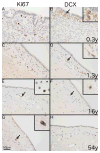
Fig. 1
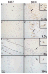
Fig. 1. Immunostaining of endogenous markers of proliferation and neurogenesis in the human SVZ
Representative photomicrographs showing differences in immunostaining for Ki67 (A, C, E and G) and DCX (B, D, F and H) with age. Immunostaining of the SVZ from a 0.3 year-old individual shows (A) numerous Ki67+ cells with a nuclear staining pattern and (B) clusters of DCX+ cells with a combination of somatic and dendritic staining. A similar pattern is shown in a 1.3 year-old individual for both Ki67 (C) and DCX (D). A 16 year-old individual shows (E) a single Ki67+ cell (black arrow) in the SVZ and (F) shows a single DCX+ cell (black arrow) with weak somatic staining. A 54 year-old individual shows (G) a single Ki67+ cell (black arrow) and (H) no DCX+ cells. 400x magnification. Scale bar = 50μm. Insets show digital enlargement of immunopositive cells.
Fig. 2
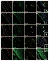
Fig. 2. Phenotype of proliferating cells in the juvenile SVZ
Confocal micrographs of the SVZ from a 1 year-old individual shows Ki67+ cells co-localising with the immature neuronal marker beta III tubulin and the neuroblast marker DCX (A–D), the transit-amplifier marker EGFR (E–H), the oligodendrocytic marker Olig2 (I–L), the microglial marker Iba1 (M–P) but not the astrocytic marker GFAP (Q–T). 400x Magnification. Scale bar = 50μm. Insets show higher magnification of Ki67+ cells.
Fig. 3
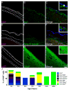
Fig. 3. Phenotype of proliferating cells in the adult SVZ
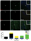
Confocal micrographs of the SVZ from a 54 year-old individual shows Ki67+ cells co-localising with the microglial marker Iba1 (A–C) but not the immature neuronal marker beta III tubulin (D–F) or the astrocytic marker GFAP (G–I). (J) A graph displaying the number of Ki67+ cells co-localising with different cell specific markers in the SVZ across a range of ages, note as quantification was performed on serial tissue sections totals could exceed or be less than 100%. (A–I) Magnification = 200x, Insets - 600x. Scale bar = 50 μm.
Fig. 4
Fig. 4. Immunostaining of endogenous markers of proliferation and neurogenesis in the human SGZ
Representative photomicrographs showing differences in immunostaining for Ki67 and DCX with age with the polymorphic layer to the right side in all images. Immunostaining shows the SGZ of a 0.3 year-old individual with (A) numerous Ki67+ cells with a nuclear staining pattern and (B) abundant DCX+ cells with predominantly somatic staining. A 1.3 year-old shows (C) a single Ki67+ cell and (D) numerous DCX+ cells with predominantly cytoplasmic staining. A16 year-old individual shows no Ki67+ cells (E) and a single DCX+ cell with dendritic and somatic staining (arrow). A 54 year-old individual shows the same pattern for both Ki67 (G) and DCX (H) as the 16 year-old individual. 400x magnification. Scale bar = 50μm.
Fig. 5
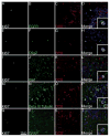
Fig. 5. Phenotype of proliferating cells in the juvenile SGZ
Confocal micrographs of the SGZ from a 1 year-old individual shows Ki67+ cells co-localising with the transit-amplifier marker EGFR (A–D), the oligodendrocytic marker Olig2 (E–H), but not the microglial marker Iba1 (I–L), the immature neuronal marker beta III tubulin (M–P) or the astrocytic marker GFAP (Q–T). 400x Magnification. Scale bar = 50 μm. Insets show digital enlargement of Ki67+ cells.
Fig. 6
Fig. 6. Phenotype of proliferating cells in the adult SGZ
Confocal micrographs of the SGZ from a 54 year-old individual shows Ki67+ cells co-localising with the microglial marker Iba1 (A–C) but not the immature neuronal marker beta III tubulin (D–F) or the astrocytic marker GFAP (G–I). (J) A graph displaying the number of Ki67+ cells co-localising with different cell specific markers in the SGZ across a range of ages. N.B. as quantification was performed on serial tissue sections totals could exceed or be less than 100%. (A–I) Magnification = 200x. Insets at 600x magnification. Scale bar = 50 μm.
Fig. 7
Fig. 7. Characterisation of the RMS
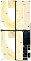
Photomicrographs show traces of the rostral migratory stream with distinctive ependymal islets extending rostrally from and confluent with the frontal horn of the lateral ventricle between the corpus callosum (CC) and head of the caudate nucleus (CN). Higher magnifications of regions corresponding to black rectangles are shown in insets. Collages of overlapping fields show DCX+ cells throughout the dorsomedial portion of the RMS in a (A) 0.2 year-old and (B) one year-old brain but only a single DCX+ cell (nuclear) in the (C) 54 year-old. (D – E) Ki67+ cells are seen in ventrolateral portion of the RMS in (D) 0.2 year-old but markedly fewer Ki67+ cells within the RMS of (E) a one year-old. (F) a confocal micrograph showing the disparity of Ki67 (red) and DCX (green) within the RMS of a 0.2 year-old individual. Orthogonal projections of z-stacks show (G) a rare Ki67+/DCX+ cells in the infant RMS whilst and (H) a Ki67+ cellsin the adult RMS co-positive for the microglia marker, Iba1.

Comment in

Similar articles

Cited by

References

    1. Falk A, Frisen J. New neurons in old brains. Ann Med. 2005;37:480–6. - PubMed
    1. Taylor CJ, Jhaveri DJ, Bartlett PF. The therapeutic potential of endogenous hippocampal stem cells for the treatment of neurological disorders. Frontiers in cellular neuroscience. 2013;7:5. - PMC - PubMed
    1. Capilla-Gonzalez V, Herranz-Perez V, Garcia-Verdugo JM. The aged brain: genesis and fate of residual progenitor cells in the subventricular zone. Frontiers in cellular neuroscience. 2015;9:365. - PMC - PubMed
    1. Mouret A, Murray K, Lledo PM. Centrifugal drive onto local inhibitory interneurons of the olfactory bulb. Ann N Y Acad Sci. 2009;1170:239–54. - PubMed
    1. Nissant A, Bardy C, Katagiri H, Murray K, Lledo PM. Adult neurogenesis promotes synaptic plasticity in the olfactory bulb. Nat Neurosci. 2009;12:728–30. - PubMed

Publication types

MeSH terms