. 2016 Oct;240(2):211-23.
doi: 10.1002/path.4769. Epub 2016 Sep 19.

Collaboration of cancer-associated fibroblasts and tumour-associated macrophages for neuroblastoma development

Affiliations

Collaboration of cancer-associated fibroblasts and tumour-associated macrophages for neuroblastoma development

Okito Hashimoto et al. J Pathol. 2016 Oct.

Abstract

Neuroblastoma is the most common extracranial solid tumour in children and is histologically classified by its Schwannian stromal cells. Although having fewer Schwannian stromal cells is generally associated with more aggressive phenotypes, the exact roles of other stromal cells (mainly macrophages and fibroblasts) are unclear. Here, we examined 41 cases of neuroblastoma using immunohistochemistry for the tumour-associated macrophage (TAM) markers CD68, CD163, and CD204, and a cancer-associated fibroblast (CAF) marker, alpha smooth muscle actin (αSMA). Each case was assigned to low/high groups on the basis of the number of TAMs or three groups on the basis of the αSMA-staining area for CAFs. Both the number of TAMs and the area of CAFs were significantly correlated with clinical stage, MYCN amplification, bone marrow metastasis, histological classification, histological type, and risk classification. Furthermore, TAM settled in the vicinity of the CAF area, suggesting their close interaction within the tumour microenvironment. We next determined the effects of conditioned medium of a neuroblastoma cell line (NBCM) on bone marrow-derived mesenchymal stem cells (BM-MSCs) and peripheral blood mononuclear cell (PBMC)-derived macrophages in vitro. The TAM markers CD163 and CD204 were significantly up-regulated in PBMC-derived macrophages treated with NBCM. The expression of αSMA by BM-MSCs was increased in NBCM-treated cells. Co-culturing with CAF-like BM-MSCs did not enhance the invasive ability but supported the proliferation of tumour cells, whereas tumour cells co-cultured with TAM-like macrophages had the opposite effect. Intriguingly, TAM-like macrophages enhanced not only the invasive abilities of tumour cells and BM-MSCs but also the proliferation of BM-MSCs. CXCL2 secreted from TAM-like macrophages plays an important role in tumour invasiveness. Taken together, these results indicate that PBMC-derived macrophages and BM-MSCs are recruited to a tumour site and activated into TAMs and CAFs, respectively, followed by the formation of favourable environments for neuroblastoma progression. © 2016 The Authors. The Journal of Pathology published by John Wiley & Sons Ltd on behalf of Pathological Society of Great Britain and Ireland.

Keywords: MSR1, macrophage scavenger receptor 1; cancer-associated fibroblast; neuroblastoma; tumour microenvironment; tumour-associated macrophage.


Figures

Figure 1
Figure 1
Immunohistochemical staining for CD68, CD163, CD204, αSMA, and h‐caldesmon in neuroblastoma samples. Two representative cases are shown. (A–F) CD68 (A), CD163 (B), CD204 (C), αSMA (D), and h‐caldesmon (E) were stained in a non‐metastatic case, stage 1. (G–L) CD68 (G), CD163 (H), CD204 (I), αSMA (J), and h‐caldesmon (K) were stained in a metastatic case, stage 4. The insets of G–L indicate high magnification views of the boxed area in each panel. Original magnification: 100×. Inset magnification: 400 × .
Figure 2
Figure 2
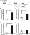
Acquisition of TAM characteristics in PBMCs treated with conditioned medium of the neuroblastoma cell line (NBCM). (A) CD14‐positive PBMCs were selected with MACS and then treated with 25 ng/ml recombinant human M‐CSF for 6 days to induce macrophage‐like differentiation, and then exposed to 50% NBCM for 2 days. (B, C) The mRNA levels of the TAM markers CD163 and CD204 in PBMC‐derived macrophages stimulated with 50% NBCM were determined by RT‐qPCR, normalized to GAPDH. (D, E) The IL10 and IL12 mRNA expression patterns in PBMC‐derived macrophages stimulated with 50% NBCM were analysed by RT‐qPCR, normalized to GAPDH expression. Values represent the mean of three experiments in duplicate and are expressed as mean ± SD (**p < 0.01).
Figure 3
Figure 3
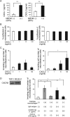
The induction of CAF markers, αSMA, and fibroblast activation protein (FAP) in human neonate fibroblasts and BM‐MSCs treated with NBCM. Fibroblasts and BM‐MSCs were exposed to 50% NBCM for 3 and 7 days. The medium was changed every 3 or 4 days. (A, B) The αSMA (ACTA2) mRNA expression in fibroblasts (A), and αSMA and FAP mRNA expression in BM‐MSCs (B) stimulated with 50% NBCM for the indicated days were determined by RT‐qPCR, normalized to GAPDH expression. Values represent the mean of three experiments in duplicate and are expressed as mean ± SD (*p < 0.05; **p < 0.01). (C) BM‐MSCs were stimulated with 50% NBCM for 7 days. After the stimulation, double immunofluorescence was performed using anti‐αSMA (red) and anti‐FAP (green). DAPI (blue) provided nuclear counterstaining. Original magnification: 200×. (D) The protein levels of αSMA, FAP, and β‐actin in BM‐MSCs stimulated with 50% NBC for the indicated days were quantified by western blotting.
Figure 4
Figure 4
Induction of growth and invasive ability under co‐culture conditions. The growth‐promoting effects of co‐culture conditions on neuroblastoma cells (A) and BM‐MSCs (B) were assessed by the MTS assay. Values represent the mean of four experiments and are expressed as mean ± SEM (*p < 0.05; **p < 0.01). Effects of the co‐culture conditions on the invasive ability of neuroblastoma cells (C), BM‐MSCs (D), and PBMCs (E) were analysed by a BioCoat Matrigel invasion chamber assay. Invading cells were counted in five randomly chosen fields. Values are the mean of four experiments and are expressed as mean ± SEM (*p < 0.05; **p < 0.01).
Figure 5
Figure 5
Effect of CXCL2 secreted from TAM‐like macrophages on tumour invasive ability. (A) CXCL1 and CXCL2 concentrations on M0 and TAM‐like macrophage supernatants were analysed by ELISA. Values represent the mean of three experiments in duplicate and are expressed as mean ± SD (**p < 0.01). (B) The growth‐promoting effects of human recombinant CXCL1 and CXCL2 in the indicated concentration on neuroblastoma cells were assessed by the MTS assay. (C) The effects of human recombinant CXCL1 and CXCL2 in the indicated concentration on the invasive ability of neuroblastoma cells were analysed by a BioCoat Matrigel invasion chamber assay. Invading cells were counted in five randomly chosen fields. (D) After expression of CXCR2 in neuroblastoma cells was confirmed by western blotting, the effect of CXCR2 Ab on the invasiveness of neuroblastoma cells co‐cultured with TAM‐like macrophages was investigated using a BioCoat Matrigel invasion chamber assay. THP‐1 cells were used as a control of CXCR2 expression. Normal mouse IgG isotype was used as a control for CXCR2 Ab. Values are the mean of three experiments and are expressed as mean ± SEM (*p < 0.05).

Comment in

References

    1. Modak S, Cheung NK. Neuroblastoma: therapeutic strategies for a clinical enigma. Cancer Treat Rev 2010; 36: 307–317. - PubMed
    1. Brodeur GM, Pritchard J, Berthold F, et al. Revisions of the international criteria for neuroblastoma diagnosis, staging, and response to treatment. J Clin Oncol 1993; 11: 1466–1477. - PubMed
    1. Li H, Fan X, Houghton J. Tumor microenvironment: the role of the tumor stroma in cancer. J Cell Biochem 2007; 101: 805–815. - PubMed
    1. Hinz B, Phan SH, Thannickal VJ, et al. Recent developments in myofibroblast biology: paradigms for connective tissue remodeling. Am J Pathol 2012; 180: 1340–1355. - PMC - PubMed
    1. Augsten M. Cancer‐associated fibroblasts as another polarized cell type of the tumor microenvironment. Front Oncol 2014; 4: 62. - PMC - PubMed

MeSH terms