. 2016 Aug 2;16(5):1366-1378.
doi: 10.1016/j.celrep.2016.06.074. Epub 2016 Jul 14.

Signal-Dependent Recruitment of BRD4 to Cardiomyocyte Super-Enhancers Is Suppressed by a MicroRNA

Affiliations

Signal-Dependent Recruitment of BRD4 to Cardiomyocyte Super-Enhancers Is Suppressed by a MicroRNA

Matthew S Stratton et al. Cell Rep. .

Abstract

BRD4 governs pathological cardiac gene expression by binding acetylated chromatin, resulting in enhanced RNA polymerase II (Pol II) phosphorylation and transcription elongation. Here, we describe a signal-dependent mechanism for the regulation of BRD4 in cardiomyocytes. BRD4 expression is suppressed by microRNA-9 (miR-9), which targets the 3' UTR of the Brd4 transcript. In response to stress stimuli, miR-9 is downregulated, leading to derepression of BRD4 and enrichment of BRD4 at long-range super-enhancers (SEs) associated with pathological cardiac genes. A miR-9 mimic represses stimulus-dependent targeting of BRD4 to SEs and blunts Pol II phosphorylation at proximal transcription start sites, without affecting BRD4 binding to SEs that control constitutively expressed cardiac genes. These findings suggest that dynamic enrichment of BRD4 at SEs genome-wide serves a crucial role in the control of stress-induced cardiac gene expression and define a miR-dependent signaling mechanism for the regulation of chromatin state and Pol II phosphorylation.


Figures

Figure 1
Figure 1. Cardiac miR-9 Expression is Suppressed During Pathological Hypertrophy
(A) Schematic representation of the rat BRD4 3′UTR with predicted microRNA (miR) binding sites indicated. (B) Hypothesis that signals for cardiac hypertrophy repress expression of a BRD4-targeting miR. (C) Expression of putative BRD4 3′ UTR binding miRs in neonatal rat ventricular myocytes (NRVMs) treated with vehicle control (Veh) or the pro-hypertrophic agonist phenylephrine (PE; 10 μM) for 48 hours. Only miR-9 expression was significantly downregulated. (D) Treatment with the histone deacetylase inhibitor (HDACi) AR-42 (500 nM) derepressed miR-9 expression in PE-treated NRVMs. miR-9 expression was also significantly downregulated in NRVMs treated with prostaglandin F2α (PGF2α; 10 μM) for 48 hours (E), in hypertrophic left ventricles (LV) of mice subjected to transverse aortic constriction (TAC), and in hypertrophic right ventricles (RV) of rats with pulmonary hypertension due to combined exposure to hypoxia and the VEGF receptor inhibitor SU5416 (F); *P<0.05 vs. vehicle or sham operated or normoxic (Nx) controls. Expression of miR-9 was significantly increased in human hearts upon mechanical unloading with a left ventricular assist device (LVAD) (G), which correlated with reduced BRD4 protein expression in the LV (H); *P<0.05 vs. Pre-LVAD. (I) Conservation of the miR-9 binding site in human, mouse and rat BRD4 3′UTR.
Figure 2
Figure 2. Cardiac BRD4 Expression is Suppressed by miR-9
(A) NRVMs were transfected with the indicated siRNAs, microRNA mimics, microRNA inhibitors or controls (Ctrl), and 48 hours post-transfection protein homogenates were analyzed by immunoblotting with the indicated antibodies; two independent anti-BRD4 antibodies were employed (Ab #1 and Ab #2), as described in the Experimental Procedures section. The miR-9 mimic reduced expression of BRD4 as efficiently as a BRD4-targeting siRNA; calnexin served as a loading control. (B) HEK293 cells were transfected with luciferase reporters fused to wildtype rat Brd4 3′ UTR or Brd4 3′UTR containing a point mutation at the miR-9 binding site. Cells were co-transfected with either miR-9 mimic or control, and luciferase activity was quantified 24 hours post-transfection; *P<0.05 vs. Ctrl mimic transfected cells.
Figure 3
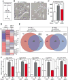
Figure 3. miR-9 and JQ1 Target Overlapping Gene Programs in Cardiomyocytes
(A) NRVMs were transfected with control mimic (Ctrl; 25 nM) or miR-9 mimic (25 nM), and 24 hours post-transfection cells were treated with vehicle (Veh) or phenylephrine (PE; 10 μM) for 48 hours. RNA was harvested for RNA-Seq. (B) NRVMs transfected with miR-9 mimic appeared smaller relative to controls, and quantitative assessment of cell size confirmed that miR-9 mimic significantly blunted PE-induced hypertrophy (C; N=24 per group; *P<0.05 vs. Ctrl). Scale bar = 100 μm. (D) Heat map summary of NRVM gene expression in the indicated treatment groups. Gene expression patterns were categorized based on responses to PE and miR-9 mimic. (E) miR-9 mimic-dependent gene expression changes were compared to changes seen with BET protein inhibition using JQ1. The Venn Diagrams indicate significant alterations in gene expression mediated by miR-9 mimic and JQ1 treatment, with both treatments blunting induction of gene expression by PE, and rescuing expression of genes that are suppressed by PE. (F) Quantitative PCR confirmed that miR-9 mimic inhibits PE-induced expression of prototypical pathological cardiac genes (N=3 per treatment group; *P<0.05).
Figure 4
Figure 4. Signal-Dependent Recruitment of BRD4 to Cardiomyocyte Gene Super-Enhancers is Blunted by miR-9
(A) NRVMs were transfected with control mimic (Ctrl; 25 nM) or miR-9 mimic (25 nM), and 24 hours post-transfection cells were treated with vehicle (Veh) or phenylephrine (PE; 10 μM) for 48 hours. Hi-Seq DNA sequencing was conducted on NRVM chromatin immunoprecipited with a BRD4-specific antibody. (B) 3,771 BRD4-enriched cardiomyocyte enhancers were identified, as defined by a distance of >500 bp from proximal promoters, and are graphed in heatmap format by treatment group. Each row shows ± 5kb centered on the BRD4 peak, with rows ordered by max BRD4 signal in each region. miR-9 mimic reduced the summed absolute BRD4 signal (rpm/bp) at enhancers, as shown below the heatmap. (C) 459 SEs, defined by BRD4 signal breadth and intensity, are plotted on the x-axis and ranked by log2 fold change upon PE treatment. (D) Box plots of BRD4 binding to SEs, categorized as PE induced, PE reduced or PE unchanged based on 1.5-fold change (induced, reduced) or less than .05-fold change (unchanged). miR-9 mimic blunted BRD4 recruitment to PE induced SEs without significantly altering binding to constitutive (unchanged) SEs. miR-9 also failed to significantly attenuate PE-mediated release of BRD4 binding from certain SEs; (*P<0.05). (E) Enhancers and SEs were ranked by BRD4 signal intensity. PE treatment dramatically enhanced BRD4 binding to SEs associated with the Nppa/Nppb and Ctgf genes, and miR-9 mimic blocked BRD4 recruitment to these SEs. BRD4 binding to SEs for the phospholamban (PLN), and Myh6/Myh7 genes were not significantly altered by PE or miR-9 mimic.
Figure 5
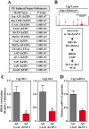
Figure 5. AP-1 Function is Required for Stimulus-Coupled Recruitment of BRD4 to Ctgf SEs
(A) Analysis of PE-inducible, BRD4-enriched SE sequences revealed enrichment of the indicated, predicted transcription factor binding sites. Binding sites for members of the AP-1 transcription factor family were overrepresented. (B) To test the hypothesis that AP-1 facilitates recruitment of BRD4 to SEs for pro-hypertrophic genes, NRVMs were infected with adenoviruses encoding dominant-negative AP-1 (Ad-dnAP-1) or β-galactosidase control (Ad-β-Gal). After 48 hours of PE treatment, sheared chromatin was subjected to anti-BRD4 ChIP, followed by PCR to quantify the presence of SE1 and SE2 of the Ctgf locus, as indicated. Expression of dnAP-1 significantly reduced the abundance of BRD4 at these SEs (C), which correlated with suppression of Ctgf mRNA expression (D); *P<0.05 vs. Ad-β-Gal.
Figure 6
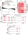
Figure 6. miR-9 Mimic Blunts Stimulus-Coupled Recruitment of BRD4 to Active Cardiomyocyte Promoters
(A) Promoters were defined in NRVMs based on Pol II and BRD4 co-occupancy. Based on Pol II enrichment, 420 BRD4-bound promoters are plotted on the x-axis and ranked by log2-fold change in BRD4 enrichment upon PE treatment (relative to control mimic + vehicle treatment). PE treatment led to BRD4 recruitment to Nppa, Nppb, and Ctgf promoters without affecting BRD4 recruitment to Myh6/Myh7 or Pln promoters. (B) BRD4-enriched active cardiomyocyte promoters are depicted in heat map format. Red intensity indicates increased BRD4 signal relative to median intensity. Stimulus-coupled recruitment of BRD4 to SEs and promoters is illustrated by the BRD4 ChIP-Seq tracks at the Nppb, Nppa, and Ctgf (C) gene loci. miR-9 mimic diminished BRD4 binding to regulatory regions for each of these genes. BRD4 binding to the SE and promoter of the constitutively expressed Pln gene (D) was unaffected by either PE or miR-9.
Figure 7
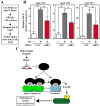
Figure 7. miR9 Suppresses Signal-Dependent Pol II Phosphorylation at Transcription Start Sites of Genes Associated With Pathologic Hypertrophy
(A) Experimental design for ChIP-PCR studies of Pol II phosphorylation at the transcription start sites (TSSs) for the Nppa, Nppb and Ctgf genes. (B) PE-mediated Pol II phosphorylation at each of these sites was significantly inhibited by miR-9; (*P<0.05). Note the correlation between changes in Pol II phosphorylation and expression of these genes (Fig. 3F). (C) A model for stimulus-dependent regulation of pathological cardiac gene expression by miR-9 and BRD4.

References

    1. Ambardekar AV, Walker JS, Walker LA, Cleveland JC, Jr, Lowes BD, Buttrick PM. Incomplete recovery of myocyte contractile function despite improvement of myocardial architecture with left ventricular assist device support. Circ Heart Fail. 2011;4:425–432. - PMC - PubMed
    1. Anand P, Brown JD, Lin CY, Qi J, Zhang R, Artero PC, Alaiti MA, Bullard J, Alazem K, Margulies KB, et al. BET bromodomains mediate transcriptional pause release in heart failure. Cell. 2013;154:569–582. - PMC - PubMed
    1. Arner E, Daub CO, Vitting-Seerup K, Andersson R, Lilje B, Drablos F, Lennartsson A, Ronnerblad M, Hrydziuszko O, Vitezic M, et al. Gene regulation. Transcribed enhancers lead waves of coordinated transcription in transitioning mammalian cells. Science. 2015;347:1010–1014. - PMC - PubMed
    1. Bisgrove DA, Mahmoudi T, Henklein P, Verdin E. Conserved P-TEFb-interacting domain of BRD4 inhibits HIV transcription. Proc Natl Acad Sci U S A. 2007;104:13690–13695. - PMC - PubMed
    1. Brown JD, Lin CY, Duan Q, Griffin G, Federation AJ, Paranal RM, Bair S, Newton G, Lichtman AH, Kung AL, et al. NF-kappaB directs dynamic super enhancer formation in inflammation and atherogenesis. Mol Cell. 2014;56:219–231. - PMC - PubMed

LinkOut - more resources