

The blood-brain barrier in Alzheimer's disease
- PMID: 27425887
- PMCID: PMC5600438
- DOI: 10.1016/j.nbd.2016.07.007
The blood-brain barrier in Alzheimer's disease
Abstract
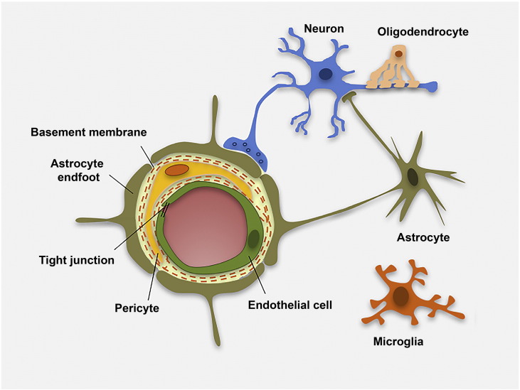
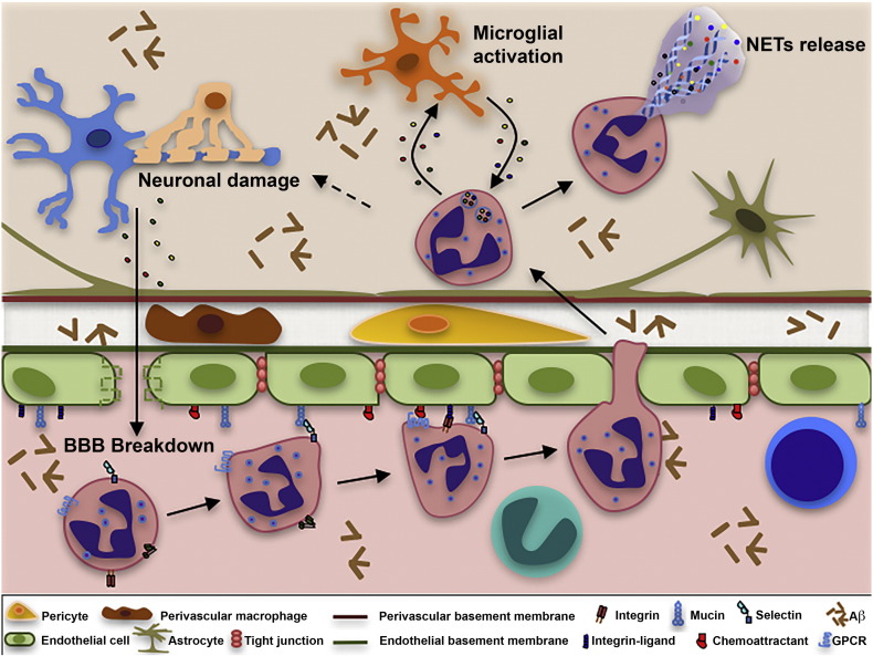
Alzheimer's disease (AD) is a chronic neurodegenerative disorder characterized by the pathological accumulation of amyloid beta (Aβ) peptides and neurofibrillary tangles containing hyperphosphorylated neuronal tau protein. AD pathology is also characterized by chronic brain inflammation, which promotes disease pathogenesis. In this context, the blood-brain barrier (BBB), a highly specialized endothelial cell membrane that lines cerebral microvessels, represents the interface between neural cells and circulating cells of the immune system. The BBB thus plays a key role in the generation and maintenance of chronic inflammation during AD. The BBB operates within the neurovascular unit (NVU), which includes clusters of glial cells, neurons and pericytes. The NVU becomes dysfunctional during AD, and each of its components may undergo functional changes that contribute to neuronal injury and cognitive deficit. In transgenic animals with AD-like pathology, recent studies have shown that circulating leukocytes migrate through the activated brain endothelium when certain adhesion molecules are expressed, penetrating into the brain parenchyma, interacting with the NVU components and potentially affecting their structural integrity and functionality. Therefore, migrating immune system cells in cerebral vessels act in concert with the modified BBB and may be integrated into the dysfunctional NVU. Notably, blocking the adhesion mechanisms controlling leukocyte-endothelial interactions inhibits both Aβ deposition and tau hyperphosphorylation, and reduces memory loss in AD models. The characterization of molecular mechanisms controlling vascular inflammation and leukocyte trafficking could therefore help to determine the basis of BBB dysfunction during AD and may lead to the development of new therapeutic approaches.
Keywords: Alzheimer's disease; Blood–brain barrier; Immune system cells; Leukocyte trafficking; Neurovascular unit; Vascular inflammation.
Copyright © 2016 The Authors. Published by Elsevier Inc. All rights reserved.
Figures
References
-
- Abbott N.J., Rönnbäck L., Hansson E. Astrocyte-endothelial interactions at the blood-brain barrier. Nat. Rev. Neurosci. 2006;7:41–53. - PubMed
-
- Abbott N.J., Patabendige A.A., Dolman D.E., Yusof S.R., Begley D.J. Structure and function of the blood-brain barrier. Neurobiol. Dis. 2010;37:13–25. - PubMed
-
- Alvarez J.I., Cayrol R., Prat A. Disruption of central nervous system barriers in multiple sclerosis. Biochim. Biophys. Acta. 2011;1812:252–264. - PubMed
Publication types
MeSH terms
Grants and funding
LinkOut - more resources
Full Text Sources
Other Literature Sources
Medical
