. 2016 Aug 8;26(15):2037-2043.
doi: 10.1016/j.cub.2016.06.002. Epub 2016 Jul 14.

The Transcription Factor ATF5 Mediates a Mammalian Mitochondrial UPR

Affiliations

The Transcription Factor ATF5 Mediates a Mammalian Mitochondrial UPR

Christopher J Fiorese et al. Curr Biol. .

Abstract

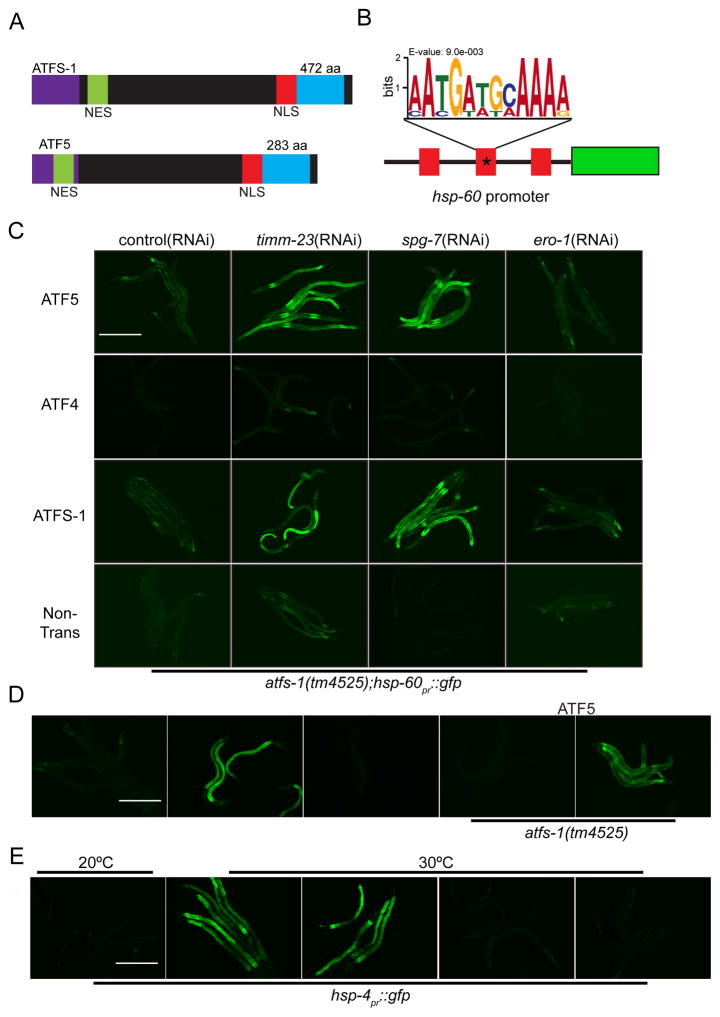
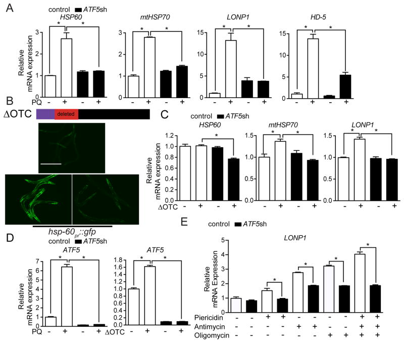
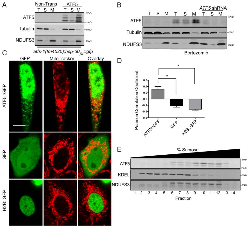
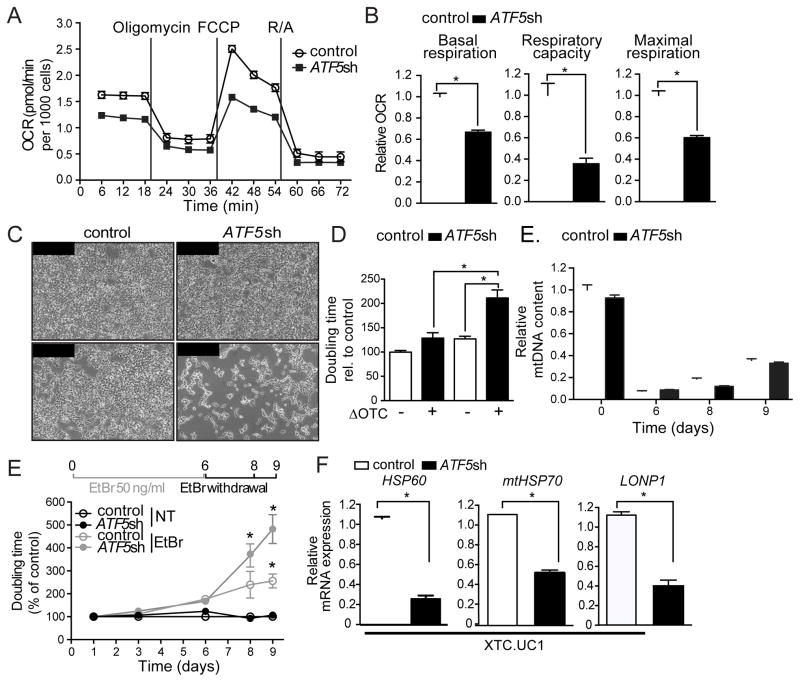
Mitochondrial dysfunction is pervasive in human pathologies such as neurodegeneration, diabetes, cancer, and pathogen infections as well as during normal aging. Cells sense and respond to mitochondrial dysfunction by activating a protective transcriptional program known as the mitochondrial unfolded protein response (UPR(mt)), which includes genes that promote mitochondrial protein homeostasis and the recovery of defective organelles [1, 2]. Work in Caenorhabditis elegans has shown that the UPR(mt) is regulated by the transcription factor ATFS-1, which is regulated by organelle partitioning. Normally, ATFS-1 accumulates within mitochondria, but during respiratory chain dysfunction, high levels of reactive oxygen species (ROS), or mitochondrial protein folding stress, a percentage of ATFS-1 accumulates in the cytosol and traffics to the nucleus where it activates the UPR(mt) [2]. While similar transcriptional responses have been described in mammals [3, 4], how the UPR(mt) is regulated remains unclear. Here, we describe a mammalian transcription factor, ATF5, which is regulated similarly to ATFS-1 and induces a similar transcriptional response. ATF5 expression can rescue UPR(mt) signaling in atfs-1-deficient worms requiring the same UPR(mt) promoter element identified in C. elegans. Furthermore, mammalian cells require ATF5 to maintain mitochondrial activity during mitochondrial stress and promote organelle recovery. Combined, these data suggest that regulation of the UPR(mt) is conserved from worms to mammals.


Figures

Figure 1
Figure 1. Expression of ATF5 rescues UPRmt activation in worms lacking ATFS-1
(A) Schematic comparing the bZip transcription factors ATFS-1 and ATF5 including the mitochondrial targeting sequence (MTS), nuclear export sequence (NES) and the nuclear localization sequence (NLS). (B) Schematic of the hsp-60pr::gfp reporter highlighting the three UPRmt elements in the promoter. The mutated element used in Figure 1D is marked with an asterisk (*). (C) Photomicrographs of atfs-1(tm4525);hsp60pr::gfp worms expressing transgenic ATF5, ATF4 or ATFS-1 and raised on control, timm-23, spg-7, or ero-1(RNAi). Scale bar, 0.5 mm. (D) Photomicrographs of wildtype and atfs-1(tm4525) worms expressing either hsp-60pr::gfp or hsp-60pr::gfp lacking a UPRmtE (*) (Figure 1B) raised on control or spg-7(RNAi). Worms in the right two panels express transgenic ATF5. Scale bar, 0.5 mm. (E) Photomicrographs of control or transgenic ATF5 expressing hsp-4pr::gfp worms, raised on control or xbp-1(RNAi) incubated at 20°C or 30°C. Scale bar, 0.5 mm. See also Figure S1.
Figure 2
Figure 2. ATF5 is required for UPRmt activation in mammalian cells
(A) Expression levels of HSP60, mtHSP70, LONP1, and HD-5 mRNA in control or ATF5 shRNA HEK 293T cells with or without paraquat (PQ) (n=3, mean ± SEM, *p<0.05). (B) Schematic showing ΔOTC construct. Photomicrographs of hsp-60pr::gfp worms raised on control or atfs-1(RNAi) with or without ΔOTC expression via the muscle-specific myo-3 promoter. Scale bar, 0.5 mm. (C) Expression levels of HSP60, mtHSP70, and LONP1 mRNA in control or ATF5 shRNA HEK 293T cells with or without ΔOTC expression (n=3, mean ± SEM, *p<0.05) (D) Expression levels of ATF5 mRNA in control or ATF5 shRNA HEK 293T cells with or without paraquat (PQ), or expressing ΔOTC (n=3, mean ± SEM, *p<0.05). (E) Expression levels of LONP1 mRNA in control or ATF5 shRNA HEK 293T cells treated with oligomycin, antimycin, piericidin, or all three inhibitors (n=3, mean ± SEM, *p<0.05). See also Figure S2.
Figure 3
Figure 3. ATF5 localizes to mitochondria and nuclei
(A) Immunoblots of lysates from control or ATF5 expressing atfs-1(tm4525);hsp60pr::gfp worms following fractionation into total lysate (T), postmitochondrial supernatant (S), and mitochondrial pellet (M). NDUFS3 serves as a mitochondrial marker and tubulin as a cytosolic marker. (B) Immunoblots of control or ATF5 shRNA HeLa cells treated with DMSO or Bortezomib and fractionated into total lysate (T), postmitochondrial supernatant (S), and mitochondrial pellet (M). (C) Photomicrographs of HeLa cells expressing either ATF5::GFP, Histone 2B::GFP (H2B::GFP), or GFP and stained with Mitotracker. Scale bar, 0.01 mm. (D) Pearson Correlation Coefficient [40] of co-localization of ATF5::GFP, Histone 2B::GFP (H2B::GFP), or GFP and MitoTracker (Figure 3C) (n=5, mean ± SEM, *p<0.05). (E) Immunoblot of mouse liver fractions following centrifugation on a sucrose gradient. Endogenous KDEL serves as an ER marker and NDUFS3 as a mitochondria marker.
Figure 4
Figure 4. ATF5 promotes proliferation and recovery from mitochondrial stress
(A–B) Oxygen consumption rates (OCR) in control or ATF5 shRNA HEK 293T cells (n=15, mean ± SEM,*p<0.05). (C–D) Photomicrographs (C) and doubling times (D) of control or ATF5 shRNA HEK 293T cells, with or without ΔOTC expression. Scale bar, 0.1 mm (n=3-7, mean ± SEM *p<0.05). (E) mtDNA quantification of control and ATF5 shRNA HEK 293T after 6 days of EtBr treatment, 4 days of withdrawal (n=3, mean ± SEM). (F) Time course of doubling times of control and ATF5 shRNA HEK 293T cells following 6 days of mtDNA depletion by EtBr treatment (n=3, mean ± SEM, *p<0.05). (G) Expression levels of HSP60, mtHSP70, or LONP1 mRNA in control or ATF5 shRNA XTC.UC1 cells (n=3, mean ± SEM, *p<0.05). See also Figure S3.

References

    1. Nargund AM, Fiorese CJ, Pellegrino MW, Deng P, Haynes CM. Mitochondrial and Nuclear Accumulation of the Transcription Factor ATFS-1 Promotes OXPHOS Recovery during the UPR. Molecular cell 2015 - PMC - PubMed
    1. Nargund AM, Pellegrino MW, Fiorese CJ, Baker BM, Haynes CM. Mitochondrial import efficiency of ATFS-1 regulates mitochondrial UPR activation. Science. 2012;337:587–590. - PMC - PubMed
    1. Wu Y, Williams EG, Dubuis S, Mottis A, Jovaisaite V, Houten SM, Argmann CA, Faridi P, Wolski W, Kutalik Z, et al. Multilayered genetic and omics dissection of mitochondrial activity in a mouse reference population. Cell. 2014;158:1415–1430. - PMC - PubMed
    1. Zhao Q, Wang J, Levichkin IV, Stasinopoulos S, Ryan MT, Hoogenraad NJ. A mitochondrial specific stress response in mammalian cells. The EMBO journal. 2002;21:4411–4419. - PMC - PubMed
    1. Pellegrino MW, Haynes CM. Mitophagy and the mitochondrial unfolded protein response in neurodegeneration and bacterial infection. BMC biology. 2015;13:22. - PMC - PubMed

MeSH terms

Substances