. 2016 Jul 18;12(7):e1005007.
doi: 10.1371/journal.pcbi.1005007. eCollection 2016 Jul.

The Integrated Role of Wnt/β-Catenin, N-Glycosylation, and E-Cadherin-Mediated Adhesion in Network Dynamics

Affiliations

The Integrated Role of Wnt/β-Catenin, N-Glycosylation, and E-Cadherin-Mediated Adhesion in Network Dynamics

Diego A Vargas et al. PLoS Comput Biol. .

Abstract

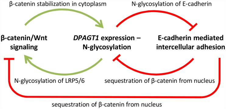
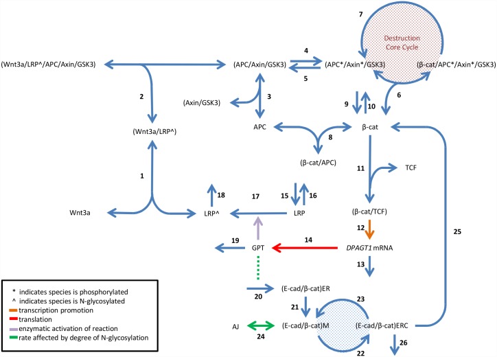
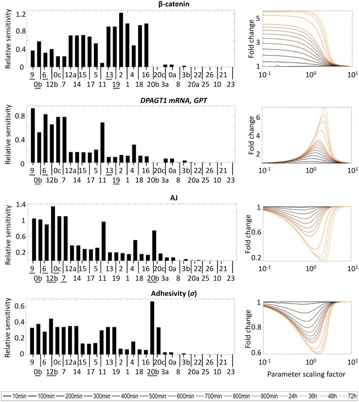
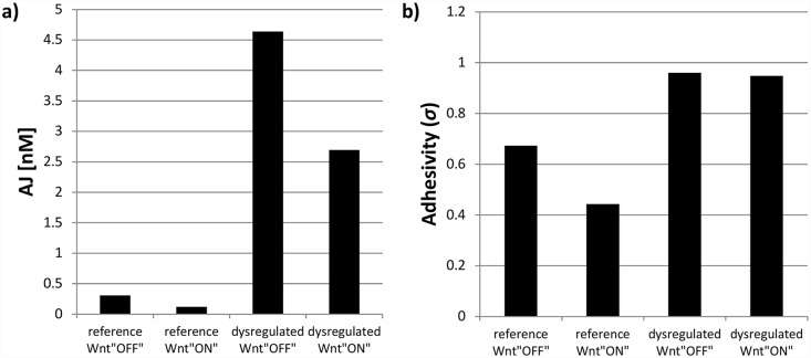
The cellular network composed of the evolutionarily conserved metabolic pathways of protein N-glycosylation, Wnt/β-catenin signaling pathway, and E-cadherin-mediated cell-cell adhesion plays pivotal roles in determining the balance between cell proliferation and intercellular adhesion during development and in maintaining homeostasis in differentiated tissues. These pathways share a highly conserved regulatory molecule, β-catenin, which functions as both a structural component of E-cadherin junctions and as a co-transcriptional activator of the Wnt/β-catenin signaling pathway, whose target is the N-glycosylation-regulating gene, DPAGT1. Whereas these pathways have been studied independently, little is known about the dynamics of their interaction. Here we present the first numerical model of this network in MDCK cells. Since the network comprises a large number of molecules with varying cell context and time-dependent levels of expression, it can give rise to a wide range of plausible cellular states that are difficult to track. Using known kinetic parameters for individual reactions in the component pathways, we have developed a theoretical framework and gained new insights into cellular regulation of the network. Specifically, we developed a mathematical model to quantify the fold-change in concentration of any molecule included in the mathematical representation of the network in response to a simulated activation of the Wnt/ β-catenin pathway with Wnt3a under different conditions. We quantified the importance of protein N-glycosylation and synthesis of the DPAGT1 encoded enzyme, GPT, in determining the abundance of cytoplasmic β-catenin. We confirmed the role of axin in β-catenin degradation. Finally, our data suggest that cell-cell adhesion is insensitive to E-cadherin recycling in the cell. We validate the model by inhibiting β-catenin-mediated activation of DPAGT1 expression and predicting changes in cytoplasmic β-catenin concentration and stability of E-cadherin junctions in response to DPAGT1 inhibition. We show the impact of pathway dysregulation through measurements of cell migration in scratch-wound assays. Collectively, our results highlight the importance of numerical analyses of cellular networks dynamics to gain insights into physiological processes and potential design of therapeutic strategies to prevent epithelial cell invasion in cancer.


Conflict of interest statement

The authors have declared that no competing interests exist.

Figures

Fig 1
Fig 1. Schematic of current notion of the relationship between Wnt/β-catenin signaling, N-glycosylation, and intercellular adhesion.
Image adapted from [25].
Fig 2
Fig 2. Reaction scheme.
Processes are numbered 1–26. Reactions 1–10 represent steps of Wnt/β-catenin signaling involved in active β-catenin regulation in the absence of Wnt3a. Reactions 11–19 represent regulation of Wnt3a binding by both genetic regulation of DPAGT1 and N-glycosylation. Reactions 20–26 represent E-cadherin dynamics and AJ formation. Abbreviations: APC, adenomatous polyposis coli; β-cat, β-catenin; E-cad, E-cadherin; ER, endoplasmic reticulum; ERC, endocytic recycling compartment; GSK3, glycogen synthase kinase 3β; LRP, lipoprotein receptor-related proteins; M, membrane; TCF, T-cell factor. Image partly adapted from [41].
Fig 3
Fig 3
(LEFT) Sensitivity to changes in individual reactions of fold-change (upon activation of Wnt/β-catenin signaling) in β-catenin, DPAGT1 mRNA and GPT, AJ, and E-cadherin adhesivity at steady-state. Reaction labels refer to numbering used in Fig 2; repeated numbers used for processes described by more than one parameter. Reaction labels along the horizontal axis are organized from left to right in order of decreasing sensitivity averaged over all molecules in network. (RIGHT) Dependence of fold-change in chosen molecules in time to changes in reaction 11 (i.e. binding equilibrium of β-catenin and TCF). The horizontal axis represents the factor by which the parameter describing binding dynamics is scaled.
Fig 4
Fig 4. Predicted values for a) adherens junctions (AJ) and b) adhesivity (σ) when numerically solving RCN model.
The four conditions simulated the experimental conditions: reference condition corresponds to physiological rate of β-catenin/TCF complex formation (i.e. equilibrium constant K11, S1 Table); dysregulated condition corresponds to ICG-001 treatment, modeled as a disruption in β-catenin/TCF complex formation (i.e. rate constant changed to 2×K11). Wnt “OFF” state is modeled with a total Wnt3a concentration WNT0 = 1 nM, and Wnt “ON” with WNT0 = 28.062 nM.
Fig 5
Fig 5. MDCK cells were treated with either conditioned media with (WCM) or without (CCM) Wnt3a, and either no inhibitor (DMSO) or ICG-001.
a) Representative immunoblots (IBs) of ABC in total cell lysates (TCL). Images were taken from a single membrane. Quantified intensity values are averages; errors bars represent standard error of the mean (SEM) (N = 4). Full IB with duplicates can be found in S3 Fig (supplemental). b) IBs for α-catenin and E-cadherin in E-cadherin immunoprecipitates (IP). CCM TLC and WCM TLC represent input, with isotype controls (IP IgG) included. Blots were quantified and normalized to the ICG-001 condition in each activation state. * represents statistically significant difference, p<0.05.
Fig 6
Fig 6. E-cadherin mobility shift from treatment of total cell lysates with glycosidases: Endoglycosidase H (EndoH) or Peptide-N-Glycosidase F (PNGaseF).
a) Immunoblots of E-cadherin from cells grown without exogenous Wnt3a (CCM). b) Immunoblots for lysate from cells grown in exogenous Wnt3a (WCM). Cells were grown in the presence of either no inhibitor (DMSO) or ICG-001.
Fig 7
Fig 7
a) Average magnitude of velocity field from PIV analysis in MDCK cell sheets and b) lateral correlation length at migrating wound edge. The four conditions correspond to treatment with: Either conditioned media with (WCM) or without (CCM) Wnt3a, and either no inhibitor (DMSO) or ICG-001. Values are the average (errors bars represent SEM) of three independent experiments for 27 time points in each (N = 81). ** and * represent statistically significant difference, p<0.005 and p<0.05 respectively.
Fig 8
Fig 8. Movement angle is the angle between expected sheet direction (i.e. wound closing or 0°) and PIV determined velocity vectors.
Color bar indicates fraction of all PIV vectors with a particular orientation. Reference condition is DMSO treated and dysregulated condition is ICG-001 treated. CCM stands for control conditioned media; WCM stands for Wnt3a conditioned media.

Similar articles

Cited by

References

    1. Masamizu Y, Ohtsuka T, Takashima Y, Nagahara H, Takenaka Y, Yoshikawa K, et al. Real-time imaging of the somite segmentation clock: revelation of unstable oscillators in the individual presomitic mesoderm cells. Proc Natl Acad Sci U S A. 2006;103: 1313–8. - PMC - PubMed
    1. Brabletz T, Jung a, Reu S, Porzner M, Hlubek F, Kunz-Schughart L a, et al. Variable beta-catenin expression in colorectal cancers indicates tumor progression driven by the tumor environment. Proc Natl Acad Sci U S A. 2001;98: 10356–61. - PMC - PubMed
    1. Näthke IS. The adenomatous polyposis coli protein: the Achilles heel of the gut epithelium. Annu Rev Cell Dev Biol. 2004;20: 337–66. - PubMed
    1. Hardy RG, Meltzer SJ, Jankowski JA. Molecular basis for risk factors Sporadic colorectal adenomas. 2000;321. - PMC - PubMed
    1. Satoh S, Daigo Y, Furukawa Y, Kato T, Miwa N, Nishiwaki T, et al. AXIN1 mutations in hepatocellular carcinomas, and growth suppression in cancer cells by virus-mediated transfer of AXIN1. Nat Genet. 2000;24: 245–50. - PubMed

Publication types

LinkOut - more resources