. 2016 Aug 1;126(8):3104-16.
doi: 10.1172/JCI85193. Epub 2016 Jul 18.

Reprogramming Müller glia via in vivo cell fusion regenerates murine photoreceptors

Reprogramming Müller glia via in vivo cell fusion regenerates murine photoreceptors

Daniela Sanges et al. J Clin Invest. .

Abstract

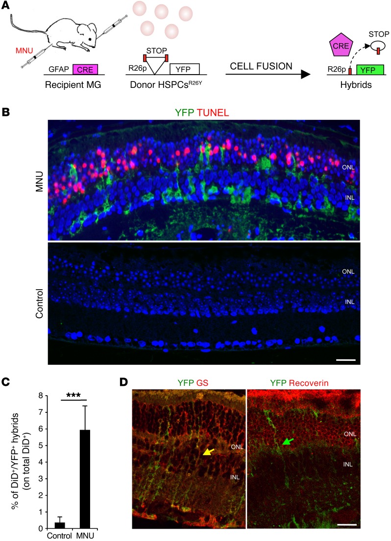
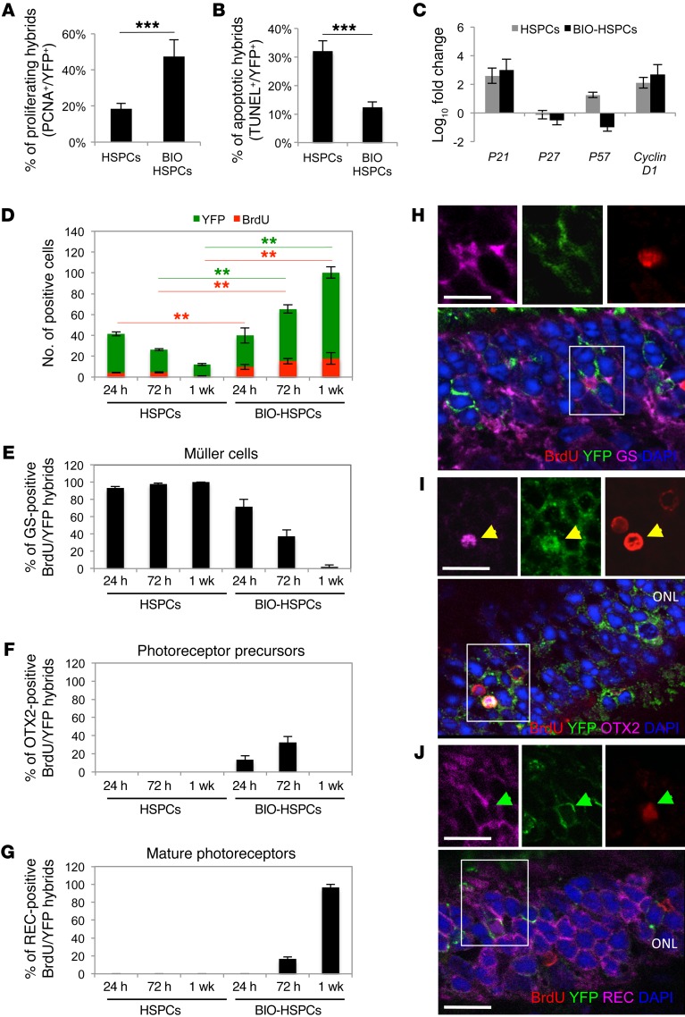
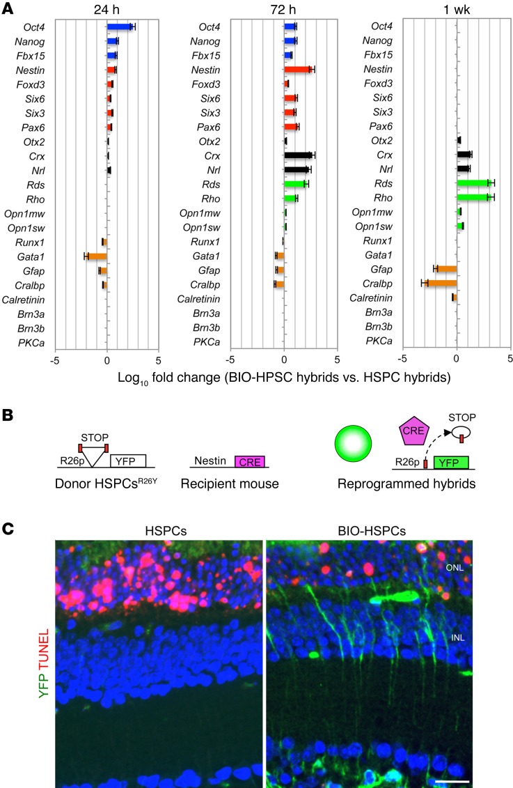
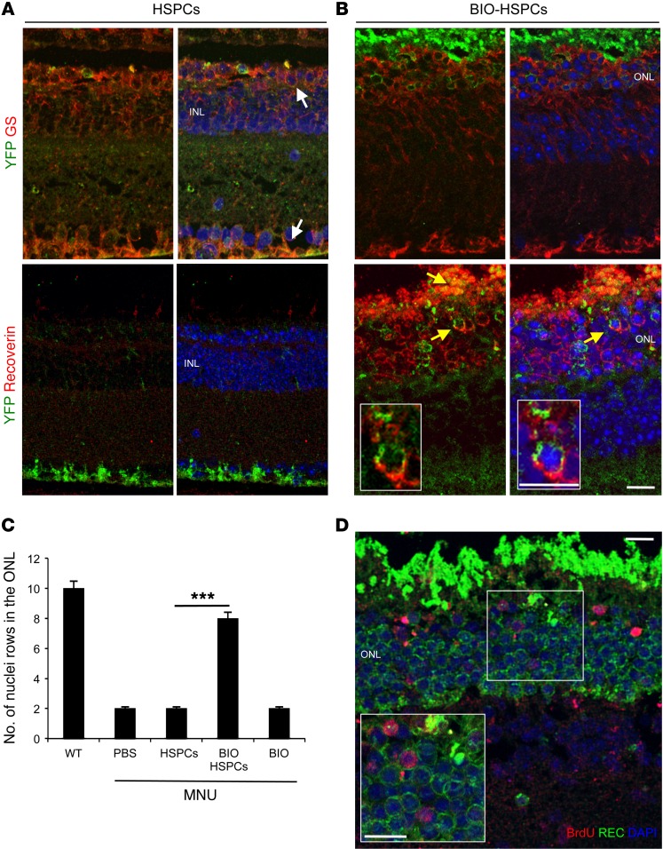
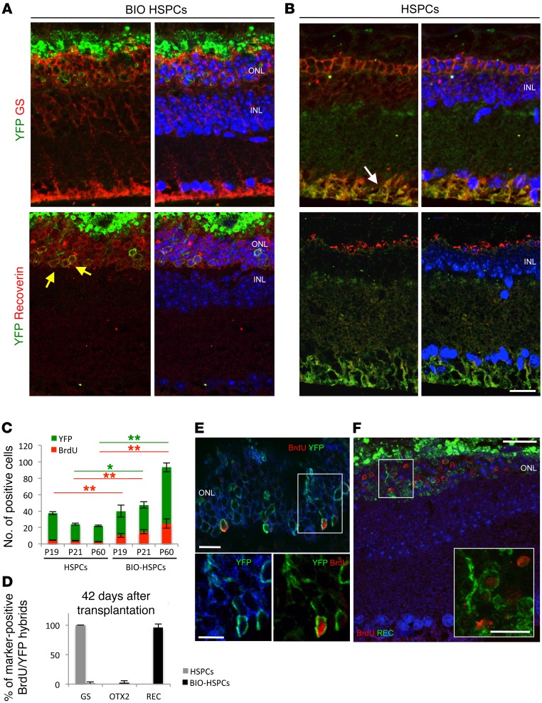
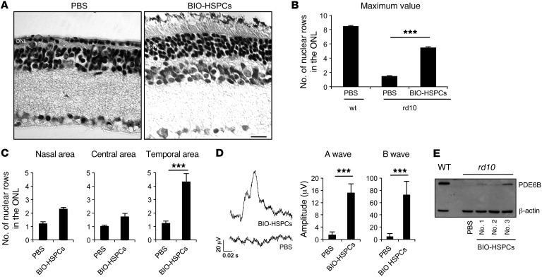
Vision impairments and blindness caused by retinitis pigmentosa result from severe neurodegeneration that leads to a loss of photoreceptors, the specialized light-sensitive neurons that enable vision. Although the mammalian nervous system is unable to replace neurons lost due to degeneration, therapeutic approaches to reprogram resident glial cells to replace retinal neurons have been proposed. Here, we demonstrate that retinal Müller glia can be reprogrammed in vivo into retinal precursors that then differentiate into photoreceptors. We transplanted hematopoietic stem and progenitor cells (HSPCs) into retinas affected by photoreceptor degeneration and observed spontaneous cell fusion events between Müller glia and the transplanted cells. Activation of Wnt signaling in the transplanted HSPCs enhanced survival and proliferation of Müller-HSPC hybrids as well as their reprogramming into intermediate photoreceptor precursors. This suggests that Wnt signaling drives the reprogrammed cells toward a photoreceptor progenitor fate. Finally, Müller-HSPC hybrids differentiated into photoreceptors. Transplantation of HSPCs with activated Wnt functionally rescued the retinal degeneration phenotype in rd10 mice, a model for inherited retinitis pigmentosa. Together, these results suggest that photoreceptors can be generated by reprogramming Müller glia and that this approach may have potential as a strategy for reversing retinal degeneration.


Figures

Figure 1
Figure 1. Transplanted HSPCs fuse with MG upon photoreceptor damage.
(A) Schematic representation of the experimental plan. Cell fusion between HSPCs isolated from LoxP-STOP-LoxP-YFP donor mice (R26Y) and recipient Gfap-Cre MG cells leads to excision of the floxed stop codon and, in turn, to the expression of YFP. (B) Representative coimmunostaining of YFP+ hybrids (green) and TUNEL+ (red) apoptotic photoreceptors on retinal sections harvested from MNU-damaged or healthy (control) Gfap-Cre eyes 12 hours after subretinal transplantation of HSPCsR26Y. YFP+ hybrids (green) derived from cell fusion are detected in MNU-damaged retinas, which show TUNEL+ photoreceptors in the ONL, but not in the undamaged eyes (control). Nuclei were counterstained with DAPI (blue). Scale bar: 20 μm. n = 3. (C) Statistical analysis of the percentage of DiD+YFP+ hybrids evaluated on the total amount of DiD-labeled HSPCsR26Y detected by FACS analysis in MNU-damaged or healthy (control) Gfap-Cre retinas 24 hours after transplantation. Data are represented as mean ± SD of 3 independent experiments. n = 3. ***P < 0.0001 by unpaired Student’s t test. (D) Representative immunostaining of YFP+ hybrids (green) also positive for the MG marker (GS, red; yellow arrows) but not for the photoreceptor marker recoverin (red; green arrow) detected 24 hours after transplantation of HSPCsR26Y in MNU-damaged Gfap-Cre retinas. Scale bar: 20 μm. n = 3.
Figure 2
Figure 2. Activation of Wnt signaling promotes proliferation and survival of hybrids.
(A and B) Percentages of proliferating (PCNA+, A) or apoptotic (TUNEL+, B) YFP+ hybrids on the total amount of YFP+ hybrids in MNU-damaged Gfap-Cre retinas 24 hours after transplantation of untreated (HSPCs) or BIO-treated (BIO-HSPCs) HSPCsR26Y. ***P < 0.0001, unpaired Student’s t test. (C) qPCR analysis of cell-cycle genes on FACS-sorted DiD+YFP+ hybrids harvested 24 hours after transplantation of untreated or BIO-treated HSPCsR26Y in MNU-damaged Gfap-Cre retinas. Data are represented as mean ± SD of log10 fold changes of gene expression in DiD+YFP+ hybrids with respect to DiD+YFP+ population-depleted retinas. n = 3. (D) Total YFP+ hybrids (green bars) that also incorporated BrdU (red bars) detected in MNU-damaged Gfap-Cre retinas 24 hours, 72 hours, and 1 week after transplantation of untreated or BIO-treated HSPCsR26Y. **P < 0.001, 2-way ANOVA and Bonferroni’s post-test. Red and green lines show the statistical significance of green (YFP) or red (BrdU) bars. (EG) Percentages of YFP+BrdU+ hybrids also positive for GS (E, Müller cells), OTX2 (F, photoreceptor progenitors), or recoverin (G, REC, mature photoreceptors) detected in MNU-damaged Gfap-Cre retinas 24 hours, 72 hours, and 1 week after transplantation of untreated or BIO-treated HSPCsR26Y. (HJ) Representative immunostainings of double YFP+ (green)/BrdU+ (red) hybrids also positive for either GS (magenta in H, white arrowheads), OTX2 (magenta in I, yellow arrowheads), or recoverin (magenta in J, green arrowheads) stainings in MNU-damaged Gfap-Cre retinas 24 hours (H), 72 hours (I), and 1 week (J) after transplantation of BIO-treated HSPCs. Nuclei were counterstained with DAPI (blue). Images on the top show higher magnification and single channels of areas in the white squares. Scale bars: 20 μm. (A, B, and DG) Values in graphs are represented as mean ± SD (n = 9).
Figure 3
Figure 3. Activation of Wnt signaling promotes reprogramming of hybrids.
(A) Gene expression analysis of pluripotent (blue bars), neural progenitor (red bars), photoreceptor progenitor (black bars), mature photoreceptor (green bars), and differentiated cell (orange bars) markers in DiD+YFP+ hybrids. The hybrids were FACS sorted 24 hours, 72 hours, and 1 week after transplantation of untreated or BIO-treated HSPCsR26Y in MNU-damaged Gfap-Cre retinas. Data represent the mean of log10 fold changes ± SEM of gene expression detected in hybrids obtained upon fusion with BIO-treated DiD-HSPCs with respect to hybrids obtained upon fusion with untreated DiD-HSPCs. n = 3. (B) Schematic representation of the experimental plan to detect dedifferentiation of retinal cells upon fusion. Reactivation of the nestin promoter in retinal cells fused with transplanted HSPCsR26Y leads to Cre expression and the consequent formation of YFP+ hybrids. (C) Representative immunostaining of YFP+ hybrids (green) and TUNEL+ apoptotic photoreceptors (red) on retinal sections obtained from MNU-damaged nestin-Cre retinas 24 hours after transplantation of untreated or BIO-treated HSPCsR26Y. Nuclei were counterstained with DAPI (blue). Scale bar: 20 μm.
Figure 4
Figure 4. MG reprogrammed upon fusion with Wnt-activated HSPCs generates new photoreceptors.
(A and B) Representative immunostainings of YFP+ (green) hybrids also immunoreactive for either GS (red, white arrows) or recoverin (red, yellow arrows) detected in MNU-damaged Gfap-Cre retinas 1 week after transplantation of untreated (A) or BIO-treated (B) HSPCsR26Y. Nuclei were counterstained with DAPI (blue). n = 3. (C) Statistical analysis of the number of photoreceptor nuclear rows in the ONL of healthy (WT) or MNU-damaged eyes 1 week after treatment with either PBS or untreated (HSPCs) or BIO-treated HSPCs (BIO-HSPCs) or BIO alone. Data are represented as mean ± SD counted in 3 different sections spanning the site of the injection for each mouse. n = 9. ***P < 0.0001, unpaired Student’s t test. (D) Representative immunodetection of BrdU-positive cells (red) also positive for the photoreceptor marker recoverin (green) in the ONL of MNU-damaged Gfap-Cre retinas 1 week after transplantation of BIO-treated HSPCsR26Y. Nuclei were counterstained with DAPI (blue). Insert represents a higher magnification of the region in the white square. (n = 3). Scale bars: 20 μm.
Figure 5
Figure 5. Regeneration of photoreceptors in rd10 mice.
(A and B) Representative immunostainings of YFP+ (green) with either GS (red, white arrow) or recoverin (red, yellow arrows) in rd10R26Y retinas 42 days after transplantation of BIO-treated (A) or untreated (B) HSPCsVav-Cre. n = 3. (C) Total number of YFP+ hybrids (green bars) that incorporated BrdU (red bars) detected in rd10R26Y eyes 24 hours (P19), 72 hours (P21), and 42 days (P60) after transplantation of untreated or BIO-treated HSPCsVav-Cre. Data are represented as mean ± SD. n = 9. **P < 0.001; *P < 0.01, 2-way ANOVA and Bonferroni’s post-test. Red and green lines show the statistical significance of the difference between the respective green (YFP) and red (BrdU) bars. (D) Percentages of double YFP+BrdU+ hybrids also positive for GS, OTX2, or recoverin stainings 42 days (P60) after transplantation of untreated (gray bars) or BIO-treated ( black bars) HSPCsVav-Cre in rd10R26Y retinas. Data are represented as mean ± SD. n = 9. (E) Representative immunodetection of BrdU+ cells (red) also positive for the photoreceptor marker recoverin (green) in the ONL of MNU-damaged rd10R26Y retinas 42 days after transplantation of BIO-treated HSPCsVav-Cre. Nuclei were counterstained with DAPI (blue). Insert shows higher magnification of the region in the white square. (F) Representative confocal image of YFP+ hybrids (green) also positive for BrdU (red) and recoverin (blue) in the ONL of rd10R26Y retinas 42 days after transplantation of BIO-treated HSPCsVav-Cre. Images at the bottom represent enlargements of the same YFP+ hybrid (green) also positive for recoverin (blue in the left panel) and for BrdU (red in the right panel) stainings. n = 3. Scale bar: 20 μm.
Figure 6
Figure 6. Functional rescue of the rd10 phenotype upon BIO-treated HSPC transplantation.
(A) Representative H&E staining of 2-month-old rd10 retinas transplanted with BIO-treated HSPCs in the right eyes or with PBS in the left eyes as control. Scale bar: 20 μm. n = 8. (B) Statistical analysis of the maximum number of nuclear rows detected in the ONL of 2-month-old mouse retinas harvested from either WT or rd10 animals transplanted at P18 with BIO-treated HSPCs or treated with vehicle alone (PBS) as control. Data are represented as mean ± SEM. n = 8. ***P < 0.0001, unpaired Student’s t test. (C) Statistical analysis of the number of ONL nuclei rows detected in the nasal, central, or temporal retinal areas of 2-month-old rd10 right retinas transplanted with BIO-treated HSPCs with respect to left control (PBS) eyes. ***P < 0.0001, unpaired Student’s t test. Nine serial sections for each mouse (n = 8) were analyzed. Counts of 8 retinas out of 21 showed increased nuclear rows and were plotted. (D) Representative ERG responses of 2-month-old rd10 mice transplanted in the right eyes with BIO-treated HSPCs (BIO-HSPCs) or with vehicle alone (PBS) in left eyes as control. Data are represented as mean ± SD (n = 8) of the A-wave and B-wave amplitudes (μV) of ERG responses from 8 out of 21 treated mice that showed increased ONL thickness upon BIO-treated HSPC transplantation. ***P < 0.0001, unpaired Student’s t test. (E) Western blotting of PDE6B protein levels in retinas harvested from either 2-month-old WT or rd10 mice treated with PBS or transplanted with BIO-treated HSPCs at P18. Three different BIO-HSPC transplanted retinas were analyzed (no. 1, no. 2, no. 3).

Similar articles

Cited by

References

    1. Blau HM, et al. Plasticity of the differentiated state. Science. 1985;230(4727):758–766. doi: 10.1126/science.2414846. - DOI - PubMed
    1. Gurdon JB, Elsdale TR, Fischberg M. Sexually mature individuals of Xenopus laevis from the transplantation of single somatic nuclei. Nature. 1958;182(4627):64–65. doi: 10.1038/182064a0. - DOI - PubMed
    1. Takahashi K, Ichisaka T, Yamanaka S. Identification of genes involved in tumor-like properties of embryonic stem cells. Methods Mol Biol. 2006;329:449–458. - PubMed
    1. Jaenisch R, Young R. Stem cells, the molecular circuitry of pluripotency and nuclear reprogramming. Cell. 2008;132(4):567–582. doi: 10.1016/j.cell.2008.01.015. - DOI - PMC - PubMed
    1. Sanges D, Cosma MP. Reprogramming cell fate to pluripotency: the decision-making signalling pathways. Int J Dev Biol. 2010;54(11–12):1575–1587. - PubMed

Publication types

MeSH terms