

Large vessel involvement by IgG4-related disease
- PMID: 27428181
- PMCID: PMC4956774
- DOI: 10.1097/MD.0000000000003344
Large vessel involvement by IgG4-related disease
Abstract
Objectives: IgG4-related disease (IgG4-RD) is an immune-mediated fibroinflammatory condition that can affect multiple organs and lead to tumefactive, tissue-destructive lesions. Reports have described inflammatory aortitis and periaortitis, the latter in the setting of retroperitoneal fibrosis (RPF), but have not distinguished adequately between these 2 manifestations. The frequency, radiologic features, and response of vascular complications to B cell depletion remain poorly defined. We describe the clinical features, radiology findings, and treatment response in a cohort of 36 patients with IgG4-RD affecting large blood vessels.
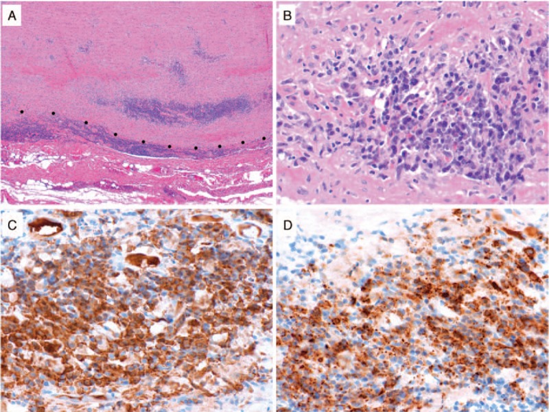
Methods: Clinical records of all patients diagnosed with IgG4-RD in our center were reviewed. All radiologic studies were reviewed. We distinguished between primary large blood vessel inflammation and secondary vascular involvement. Primary involvement was defined as inflammation in the blood vessel wall as a principal focus of disease. Secondary vascular involvement was defined as disease caused by the effects of adjacent inflammation on the blood vessel wall.
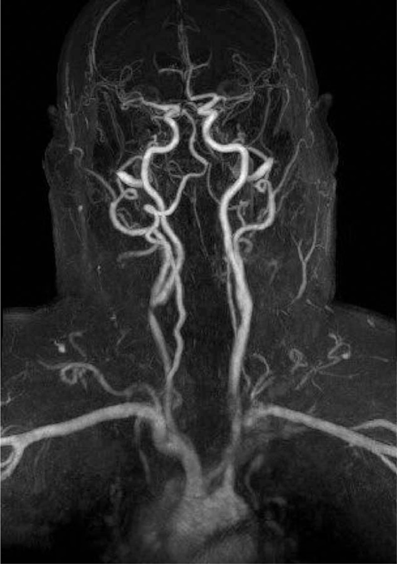
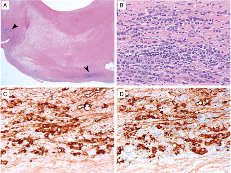
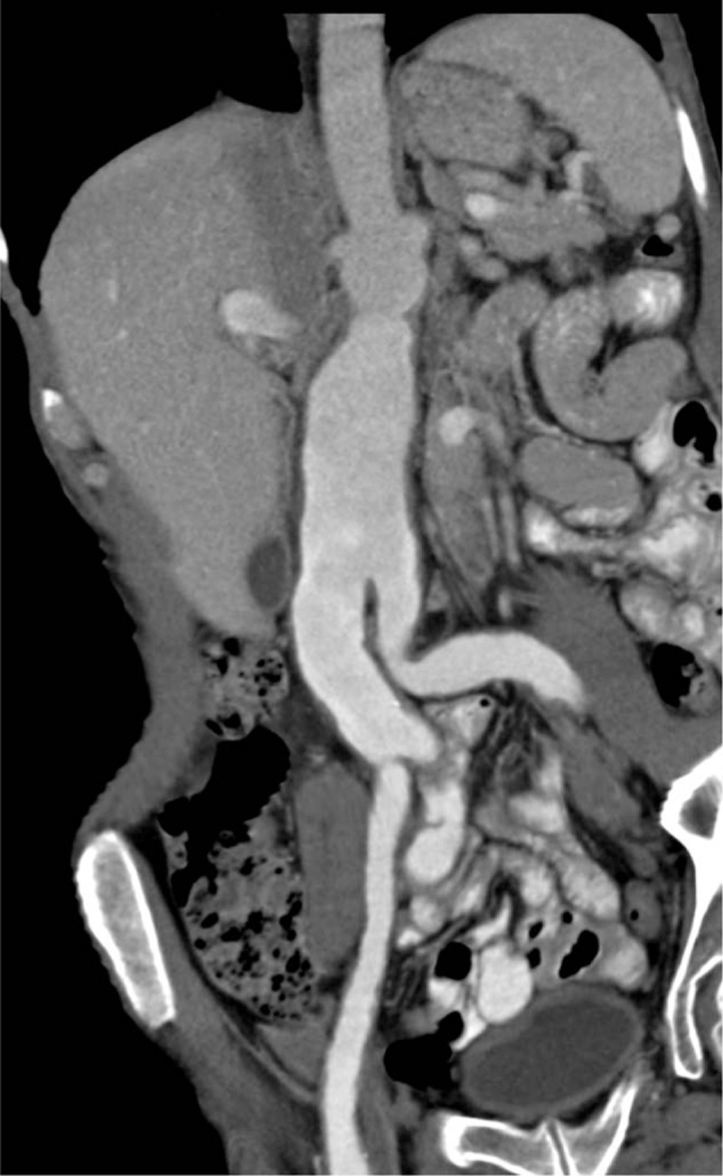
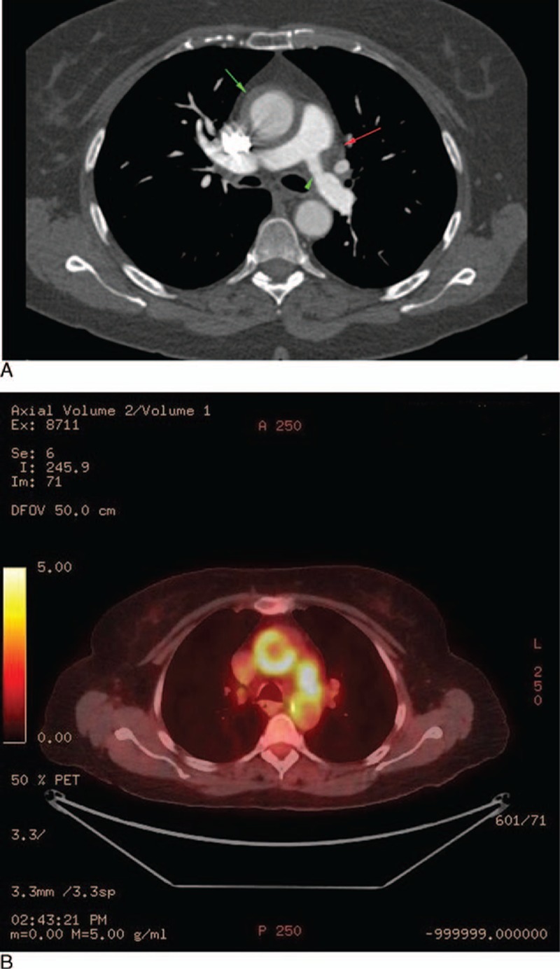
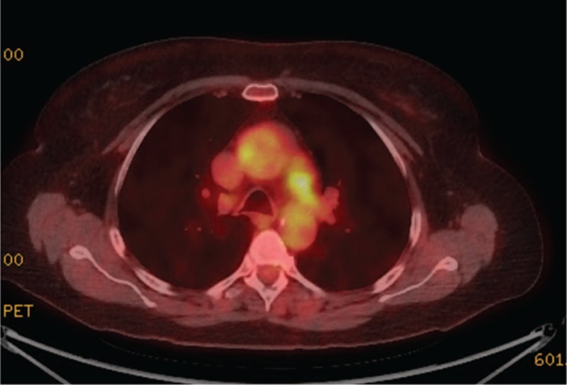
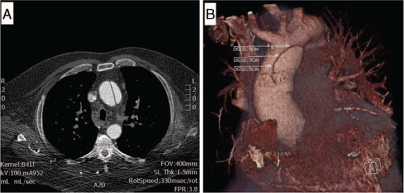
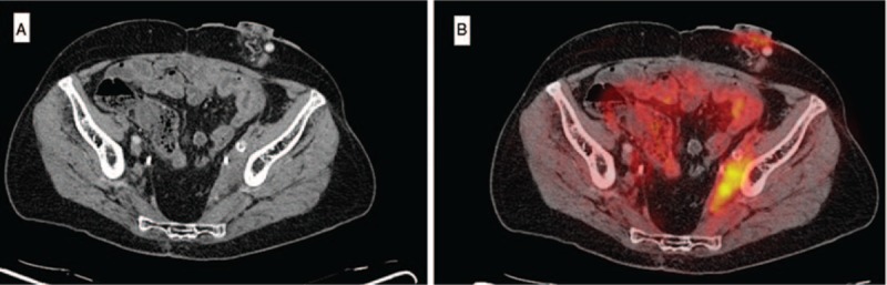
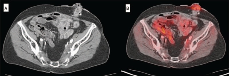
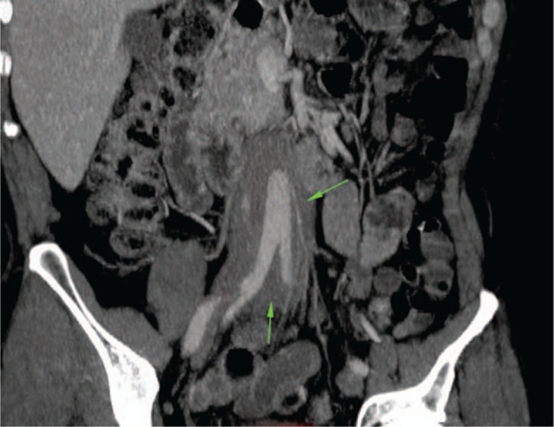
Results: Of the 160 IgG4-RD patients in this cohort, 36 (22.5%) had large-vessel involvement. The mean age at disease onset of the patients with large-vessel IgG4-RD was 54.6 years. Twenty-eight patients (78%) were male and 8 (22%) were female. Thirteen patients (36%) had primary IgG4-related vasculitis and aortitis with aneurysm formation comprised the most common manifestation. This affected 5.6% of the entire IgG4-RD cohort and was observed in the thoracic aorta in 8 patients, the abdominal aorta in 4, and both the thoracic and abdominal aorta in 3. Three of these aneurysms were complicated by aortic dissection or contained perforation. Periaortitis secondary to RPF accounted for 27 of 29 patients (93%) of secondary vascular involvement by IgG4-RD. Only 5 patients demonstrated evidence of both primary and secondary blood vessel involvement. Of those treated with rituximab, a majority responded positively.
Conclusions: IgG4-RD is a distinctive, unique, and treatable cause of large-vessel vasculitis. It can also involve blood vessels secondary to perivascular tumefactive lesions. The most common manifestation of IgG4-related vasculitis is aortitis with aneurysm formation. The most common secondary vascular manifestation is periaortitis with relative sparing of the aortic wall. Both primary vasculitis and secondary vascular involvement respond well to B cell depletion therapy.
Conflict of interest statement
The authors have no funding and conflicts of interest to disclose.
Figures

References
-
- Stone JH, Zen Y, Deshpande V. IgG4-related disease. N Engl J Med 2012; 366:539–551. - PubMed
-
- Kamisawa T, Egawa N, Nakajima H. Autoimmune pancreatitis is a systemic autoimmune disease. Am J Gastroenterol 2003; 98:2811–2812. - PubMed
-
- Weber SM, Dubukcu-Dimopulo O, Palesty A, et al. Lymphoplasmacytic sclerosing pancreatitis: inflammatory mimic of pancreatic carcinoma. J Gastrointest Surg 2003; 7:129–139. - PubMed
-
- Kasashima S, Zen Y, Kawashima A, et al. Inflammatory abdominal aortic aneurysm: close relationship to IgG4-related peri-aortitis. Am J Surg Pathol 2008; 32:197–204. - PubMed
MeSH terms
Substances
Grants and funding
LinkOut - more resources
Full Text Sources
Other Literature Sources
Medical
Miscellaneous
