. 2016 Jul 18;11(7):e0158847.
doi: 10.1371/journal.pone.0158847. eCollection 2016.

Differences in Number of Midbrain Dopamine Neurons Associated with Summer and Winter Photoperiods in Humans

Affiliations

Differences in Number of Midbrain Dopamine Neurons Associated with Summer and Winter Photoperiods in Humans

Tim D Aumann et al. PLoS One. .

Abstract

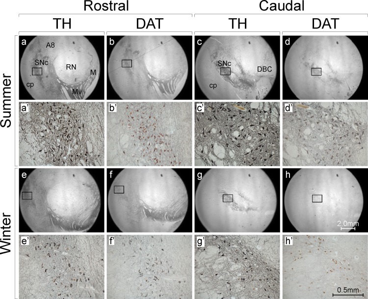
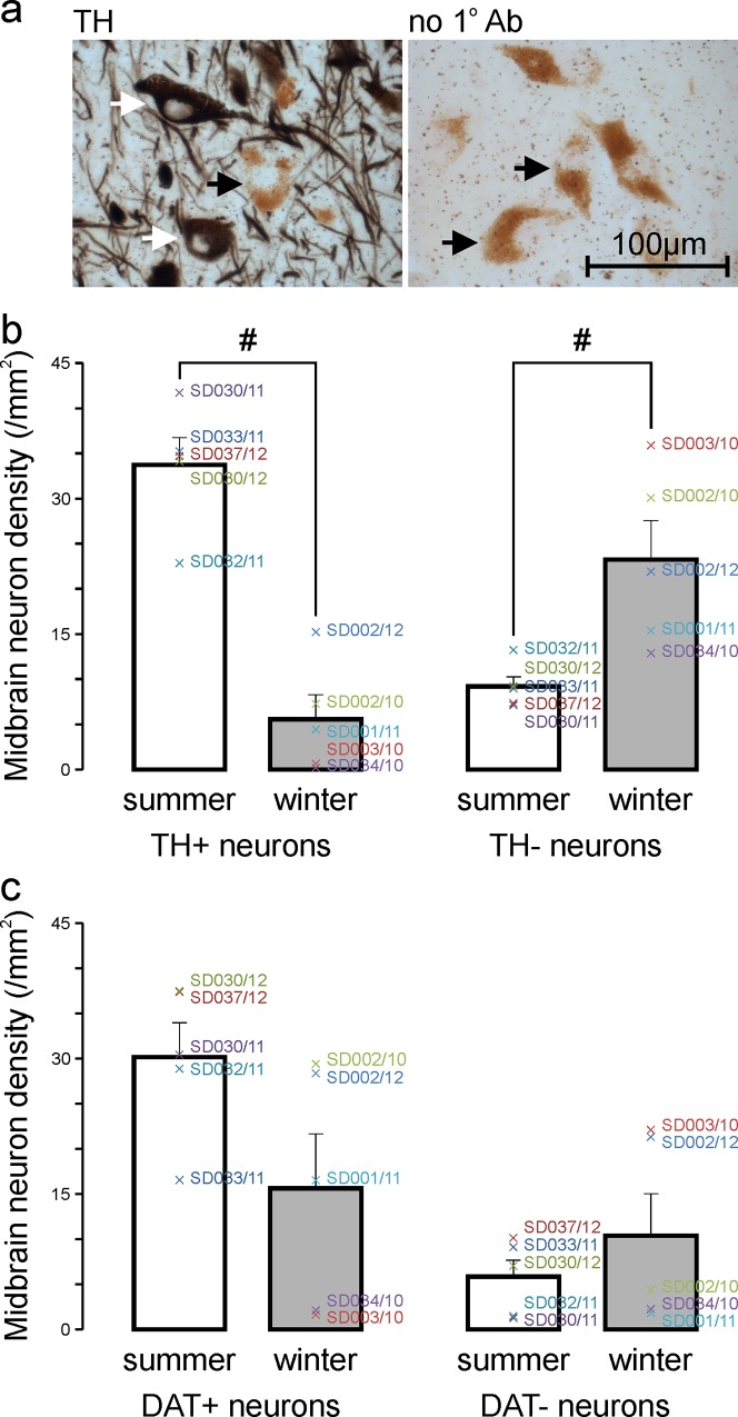
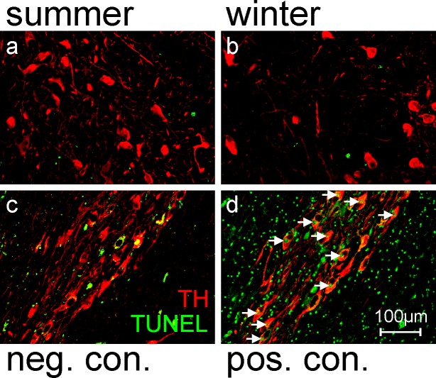
Recent evidence indicates the number of dopaminergic neurons in the adult rodent hypothalamus and midbrain is regulated by environmental cues, including photoperiod, and that this occurs via up- or down-regulation of expression of genes and proteins that are important for dopamine (DA) synthesis in extant neurons ('DA neurotransmitter switching'). If the same occurs in humans, it may have implications for neurological symptoms associated with DA imbalances. Here we tested whether there are differences in the number of tyrosine hydroxylase (TH, the rate-limiting enzyme in DA synthesis) and DA transporter (DAT) immunoreactive neurons in the midbrain of people who died in summer (long-day photoperiod, n = 5) versus winter (short-day photoperiod, n = 5). TH and DAT immunoreactivity in neurons and their processes was qualitatively higher in summer compared with winter. The density of TH immunopositive (TH+) neurons was significantly (~6-fold) higher whereas the density of TH immunonegative (TH-) neurons was significantly (~2.5-fold) lower in summer compared with winter. The density of total neurons (TH+ and TH- combined) was not different. The density of DAT+ neurons was ~2-fold higher whereas the density of DAT- neurons was ~2-fold lower in summer compared with winter, although these differences were not statistically significant. In contrast, midbrain nuclear volume, the density of supposed glia (small TH- cells), and the amount of TUNEL staining were the same in summer compared with winter. This study provides the first evidence of an association between environmental stimuli (photoperiod) and the number of midbrain DA neurons in humans, and suggests DA neurotransmitter switching underlies this association.


Conflict of interest statement

Competing Interests: The authors have declared that no competing interests exist.

Figures

Fig 1
Fig 1. Midbrain tyrosine hydroxylase (TH) and dopamine transporter (DAT) immunoreactivity are higher in summer than in winter.
Shown are photomicrographs of immunoreacted (black DAB reaction product) and Nissl-counterstained (red) human post-mortem sections at rostral (a,b,e,f) and caudal (c,d,g,h) levels of the midbrain in subjects who died in summer (a,b = SD033/11; c,d = SD030/11) and winter (e,f = SD002/10; g,h = SD034/10). The regions outlined with squares in a-h are shown at higher magnification in a’-h’. Note the increased density of immunoreactive (black) cell bodies and neural processes in summer compared with winter, more obvious for TH than DAT. A8 = dopaminergic group A8; cp = cerebral peduncle; DBC = decussation of the brachium conjunctivum; M = medial dopaminergic group; Mv = medioventral dopaminergic group; RN = red nucleus; SNc = substantia nigra pars compacta
Fig 2
Fig 2. The density of tyrosine hydroxylase immunoreactive (TH+) midbrain neurons is higher in summer than in winter.
a Cell-type classifications were made during cell density measurements. Only large (neuronal) cells with a visible nucleus were counted. TH+ neurons contained black TH immunohistochemistry reaction product (e.g. white arrows), whereas TH- neurons (e.g. black arrows) had no black reaction product but contained brown clusters of neuromelanin granules around the nucleus and/or Nissl counterstain (red staining). The photomicrograph on the left is of a section processed with the primary TH antibody, the photomicrograph on the right is of a negative control section processed without the primary TH antibody (no 1° Ab). b Mean ± SE density of midbrain TH+ (left) and TH- (right) neurons in the summer (white bar) and winter (gray bar) groups (n = 5 in each case). Individual subject means are indicated by colored crosses. TH+ neuron density is significantly higher in summer compared with winter (#, p<0.017, unpaired two-tailed t-test with correction for multiple comparisons), and TH- neuron density is significantly lower in summer compared with winter (#, p<0.017, unpaired two-tailed t-test with correction for multiple comparisons). c Mean ± SE density of midbrain DAT+ (left) and DAT- (right) neurons in the summer (white bar) and winter (gray bar) groups (n = 5 in each case). Individual subject means are indicated by colored crosses. Although seasonal differences in both DAT+ and DAT- neurons show the same trends as TH+ and TH- neurons, the differences are not statistically significant (DAT+, p>0.017; DAT-, p>0.017, unpaired two-tailed t-tests with correction for multiple comparisons)
Fig 3
Fig 3. TUNEL staining in midbrain DA nuclei is the same in summer and winter, suggesting apoptosis is not the cause of seasonal differences in TH+ and TH- neurons.
a & b Photomicrographs of sections through approximately the same area of midbrain in a summer (a) and winter (b) subject. These sections have been immunoreacted for TH (red) and stained for TUNEL (green). Note the increased density of TH+ neurons in summer but no difference in the amount of TUNEL staining between summer and winter. c & d Photomicrographs of TUNEL negative (c) and TUNEL positive (d) control sections. Both have been immunoreacted for TH (red), but the TUNEL negative control section was processed without the enzyme component of the kit, and the TUNEL positive control section was treated with DNAse prior to the TUNEL reaction. Note the lack of TUNEL+ nuclei in (c) and the large number of TUNEL+ nuclei in (d), including large TUNEL+ & TH+ nuclei (white arrows) and small TUNEL+ nuclei that are presumably glial cells

Similar articles

Cited by

References

    1. Dulcis D, Jamshidi P, Leutgeb S, Spitzer NC (2013) Neurotransmitter switching in the adult brain regulates behavior. Science 340: 449–453. 10.1126/science.1234152 - DOI - PubMed
    1. Romeo S, Viaggi C, Di Camillo D, Willis AW, Lozzi L, et al. (2013) Bright light exposure reduces TH-positive dopamine neurons: implications of light pollution in Parkinson's disease epidemiology. Sci Rep 3: 1395 10.1038/srep01395 - DOI - PMC - PubMed
    1. Aumann TD, Tomas D, Horne MK (2013) Environmental and behavioral modulation of the number of substantia nigra dopamine neurons in adult mice. Brain Behav 3: 617–625. 10.1002/brb3.163 - DOI - PMC - PubMed
    1. Tomas D, Prijanto AH, Burrows EL, Hannan AJ, Horne MK, et al. (2015) Environmental modulations of the number of midbrain dopamine neurons in adult mice. J Vis Exp. - PMC - PubMed
    1. Aumann TD, Egan K, Lim J, Boon WC, Bye CR, et al. (2011) Neuronal activity regulates expression of tyrosine hydroxylase in adult mouse substantia nigra pars compacta neurons. J Neurochem 116: 646–658. 10.1111/j.1471-4159.2010.07151.x - DOI - PubMed