. 2016 Jun:8:117-131.
doi: 10.1016/j.ebiom.2016.04.032. Epub 2016 Apr 29.

Small Molecule Inhibitor of CBFβ-RUNX Binding for RUNX Transcription Factor Driven Cancers

Affiliations

Small Molecule Inhibitor of CBFβ-RUNX Binding for RUNX Transcription Factor Driven Cancers

Anuradha Illendula et al. EBioMedicine. 2016 Jun.

Erratum in

Abstract

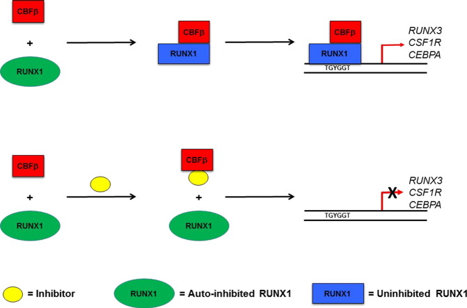
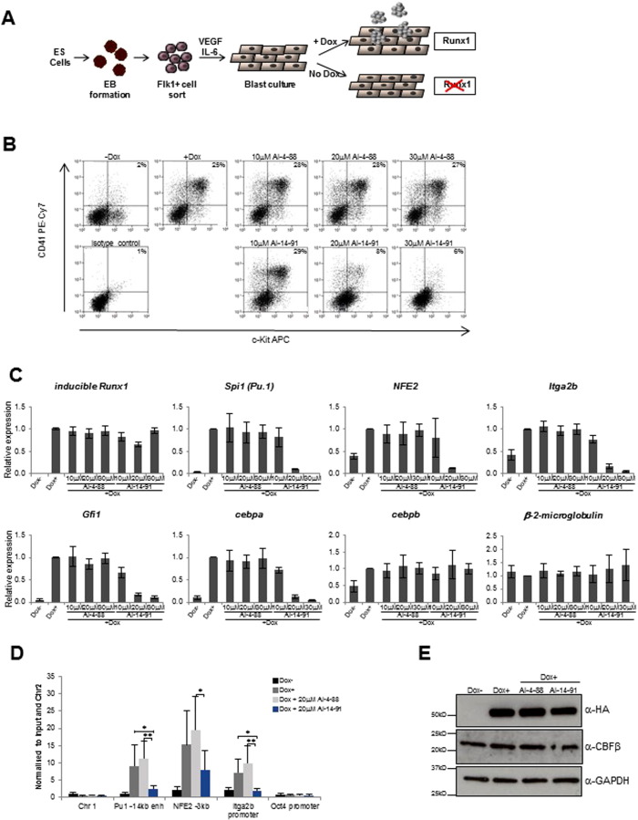
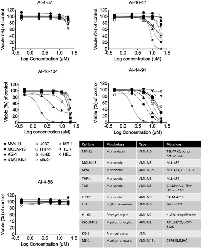
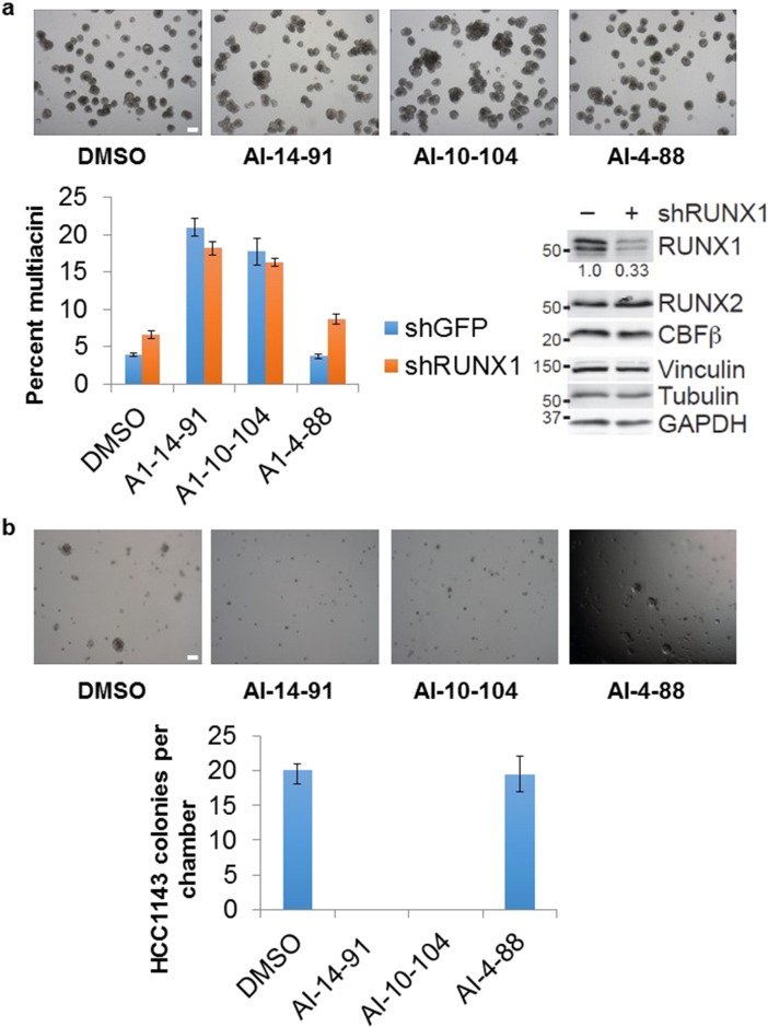
Transcription factors have traditionally been viewed with skepticism as viable drug targets, but they offer the potential for completely novel mechanisms of action that could more effectively address the stem cell like properties, such as self-renewal and chemo-resistance, that lead to the failure of traditional chemotherapy approaches. Core binding factor is a heterodimeric transcription factor comprised of one of 3 RUNX proteins (RUNX1-3) and a CBFβ binding partner. CBFβ enhances DNA binding of RUNX subunits by relieving auto-inhibition. Both RUNX1 and CBFβ are frequently mutated in human leukemia. More recently, RUNX proteins have been shown to be key players in epithelial cancers, suggesting the targeting of this pathway could have broad utility. In order to test this, we developed small molecules which bind to CBFβ and inhibit its binding to RUNX. Treatment with these inhibitors reduces binding of RUNX1 to target genes, alters the expression of RUNX1 target genes, and impacts cell survival and differentiation. These inhibitors show efficacy against leukemia cells as well as basal-like (triple-negative) breast cancer cells. These inhibitors provide effective tools to probe the utility of targeting RUNX transcription factor function in other cancers.

Keywords: CBFβ; Leukemia; PPI; RUNX; Transcription factor inhibitor; Triple negative breast cancer.


Figures

Image 10
Graphical abstract
Fig. 1
Fig. 1
Identification of pharmacophore. A. FRET assay results for AI-4-57, AI-10-104, and AI-14-91. Cerulean-Runt domain and Venus-CBFβ concentration was 100 nM. X-axis indicates compound concentration and y-axis is ratio between the emission intensities at 525 and 474 nm. Experiments were performed in duplicate. Error bars represent average ± standard deviation. The average data points were fit using Origin. B. Schemes 1–3 illustrate the targets of modifications to AI-4-57 as well as compounds synthesized and assayed to delineate the active pharmacophore. C. Results of STD NMR experiment with AI-4-57 (2 mM) and CBFβ (200 μM). Bottom panel shows 1D NMR spectrum of AI-4-57. Upper 2 panels show STD difference spectra (off-resonance (70 ppm) saturation spectrum minus on-resonance (0.4 ppm) CBFβ saturation spectrum) with saturation times of 500 and 2000 ms.
Fig. 2
Fig. 2
GLIDE based docking of inhibitors to CBFβ. A. Surface representation of the binding pocket on CBFβ (grey) with AI-4-57 (blue) bound as determined using GLIDE, all overlayed on a ribbon representation of the structure of CBFβ. B. Surface representation of the binding pocket on CBFβ with AI-4-57 bound colored according to partial charge (red, negative; blue, positive; green, neutral) in same orientation as in A. C. Schematic showing the identity of the residues making contact with AI-4-57. D. Ribbon representation of the structure of CBFβ (blue) with AI-4-57 bound. The sidechains of R90 and K111 are displayed and colored red. E. Overlays of selected regions of 15N-1H HSQC spectra of CBFβ alone and CBFβ + AI-4-57 for wildtype, R90E, and K111E CBFβ proteins. Resonances for CBFβ alone are in blue and those for CBFβ + AI-4-57 are in red.
Fig. 3
Fig. 3
Effects of CBFβ inhibitors on protein chemical shifts and backbone dynamics measured using NMR spectroscopy. A. Structure of the CBFβ (green) – Runt domain (magenta) heterodimer with a surface representation colored in red for the residues in the AI-4-57 binding pocket which undergo chemical shift changes upon binding. The orientation of AI-4-57 is that deduced from GLIDE docking. B. Structure of the CBFβ-Runt domain heterodimer with residues on the Runt domain binding interface which display chemical shift changes upon AI-4-57 binding having their surface colored in red. Residues in the Runt domain which make contact with these CBFβ residues are displayed as magenta colored surface representations. C. Plot of the difference in the measured 15N backbone R1 ∗ R2 values between CBFβ + AI-4-57 and CBFβ alone. D. Structure of the CBFβ-Runt domain heterodimer with the residues in the AI-4-57 binding pocket which display changes in R1 ∗ R2 upon AI-4-57 binding indicated as a surface representation. Residues showing increased ps-ns timescale motion are colored red and those showing increased μs-ms timescale motion are colored blue. E. Structure of the CBFβ-Runt domain heterodimer with residues on the Runt domain binding interface on CBFβ which show changes in R1 ∗ R2 upon AI-4-57 indicated as surface representations. Red indicates increased ps-ns timescale motion and blue indicates increased μs-ms timescale motion. Residues in the Runt domain which make contact with these residues are indicated as a surface representation and colored magenta. The identities of the residues involved are indicated in green for CBFβ and in magenta for the Runt domain.
Fig. 4
Fig. 4
CBFβ inhibitors reduce CBFβ binding to RUNX1 in cells. Co-immunoprecipitation assays of lysates from acute myeloid leukemia SEM cells treated with AI-4-88, AI-10-47, AI-10-104, AI-12-126 and AI-14-91 at 10 μM for 6 h. RUNX1-bound CBFβ protein levels and immunoprecipitated RUNX1 are shown on the top panel. Quantification of RUNX1 bound CBFβ is shown on the bottom graph. Protein levels were normalized to RUNX1 and depicted relative to DMSO control.
Fig. 5
Fig. 5
Effects of CBFβ inhibitors on RUNX1 occupancy and target gene expression. A. Schematic diagram representing the inducible Runx1 (iRx) ES cell differentiation system. ES cells were allowed to form embryoid bodies (EB) in IVD culture media and hemangioblast Flk1 + ve cells were sorted and seeded into blast culture media and cultured for 40 h. In the absence of doxycycline (− dox) RUNX1 is absent and cells cannot progress from the HE1 stage of development. In the presence of doxycycline (+ dox) Runx1 is expressed and differentiation progresses, resulting in the formation of CD41 + ve/KIT + ve progenitors. B. Representative FACS histograms showing staining of the surface markers CD41 and KIT in cells treated with the compounds and dox as indicated. The percentage of cells expressing KIT and CD41 is reduced by treatment with AI-14-91 but not with the control compound AI-4-88. C. Gene expression analysis of inhibitor treated cells using RNA prepared from cells treated with and without dox and cells treated with 10 μM, 20 μM and 30 μM AI-14-91 or AI-4-88 in the presence of dox. Relative expression of genes is shown, normalized to GAPDH and using + dox as the calibrator. Error bars represent standard deviation where n = 3. D. ChIP analysis of HA-Runx1 binding following Runx1 induction by dox and treatment with inhibitor or control compound. ChIP with an anti-HA antibody recognizing HA tagged Runx1 was used to identify binding at selected amplicons and enrichment normalized to input and the Chr2 control is shown. Error bars represent standard deviation where n = 5. A one-way ANOVA test was used to analyze variance in HA enrichment values between the inhibitor and control compound, or between inhibitor and + dox treatment, * denotes p < 0.05 and ** denotes p < 0.01. E. Representative western blots showing levels of inducible Runx1 and CBFβ protein in the cell populations. Where indicated, cells were treated with 20 μM inhibitor or control compound.
Fig. 6
Fig. 6
Effects of CBFβ inhibitors on the viability of a panel of leukemia cell lines. Percent viability for 11 leukemia cell lines after 48 h of treatment with the indicated compounds. The viability is represented as the percent DAPI negative cells relative to DMSO control. Each symbol/color represents an individual cell line. The symbol represents the mean of independent replicates and the error bars represent the S.E.M. The table indicates the morphology, type of leukemia, and known mutations for each of the 11 cell lines.
Fig. 7
Fig. 7
Effects of CBFβ inhibitors on acini formation by MCF10A-5E cells and viability of basal-like breast cancer cell line HC1143. CBFβ inhibitors alter acinar morphogenesis of basal-like breast epithelial cells and block survival of basal-like breast cancer cells in 3D organotypic culture. A. MCF10A-5E cells stably expressing shRUNX1 or shGFP control were grown in 3D culture for 10–11 days with the indicated compounds at 1 μM concentration and imaged by brightfield microscopy. The total number of multiacini per chamber was counted and scaled to the shGFP DMSO control. Data are shown as the mean ± s.e.m. of eight independent cultures. Quantitative immunoblotting for RUNX1, RUNX2, CBFβ, vinculin, tubulin, and GAPDH is shown to the right. B. HCC1143 cells were grown in 3D culture for 18 days with the indicated compounds at 1 μM concentration and imaged by brightfield microscopy. The total number of proliferating colonies per chamber was counted, and data are shown as the mean ± s.e.m. of four independent cultures. Scale bar is 200 μm.

References

    1. Adya N., Castilla L.H., Liu P.P. Function of CBFbeta/Bro proteins. Semin. Cell Dev. Biol. 2000;11:361–368. - PubMed
    1. Arkin M.R., Tang Y., Wells J.A. Small-molecule inhibitors of protein-protein interactions: progressing toward the reality. Chem. Biol. 2014;21:1102–1114. - PMC - PubMed
    1. Arkin M.R., Tang Y., Wells J.A. Small-molecule inhibitors of protein-protein interactions: progressing toward the reality. Chem. Biol. 2014;21:1102–1114. - PMC - PubMed
    1. Arkin M.R., Whitty A. The road less traveled: modulating signal transduction enzymes by inhibiting their protein-protein interactions. Curr. Opin. Chem. Biol. 2009;13:284–290. - PubMed
    1. Bakshi R., Hassan M.Q., Pratap J., Lian J.B., Montecino M.A., Van Wijnen A.J., Stein J.L., Imbalzano A.N., Stein G.S. The human SWI/SNF complex associates with RUNX1 to control transcription of hematopoietic target genes. J. Cell. Physiol. 2010;225:569–576. - PMC - PubMed

MeSH terms

Substances