. 2016 Sep;12(9):717-23.
doi: 10.1038/nchembio.2128. Epub 2016 Jul 18.

Targeted inhibition of oncogenic miR-21 maturation with designed RNA-binding proteins

Affiliations

Targeted inhibition of oncogenic miR-21 maturation with designed RNA-binding proteins

Yu Chen et al. Nat Chem Biol. 2016 Sep.

Abstract

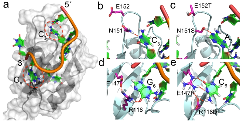
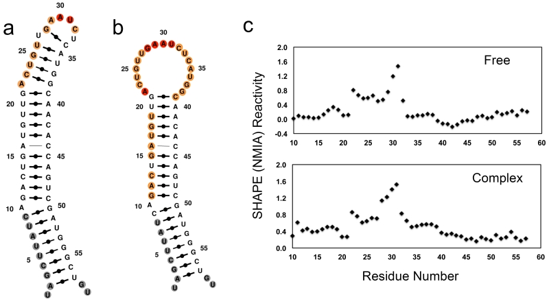
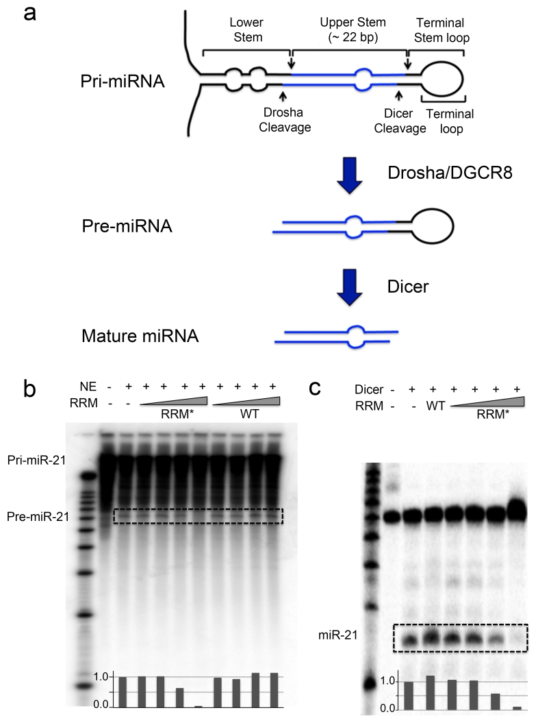
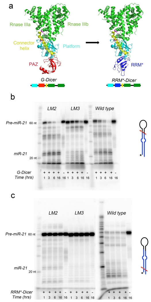
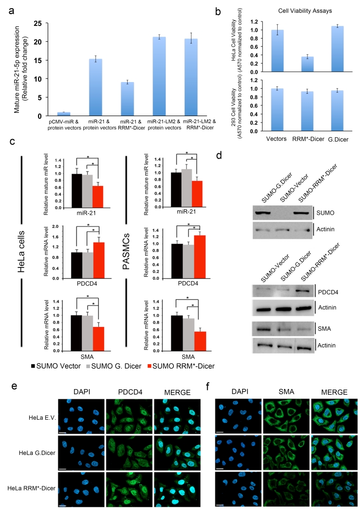
The RNA recognition motif (RRM) is the largest family of eukaryotic RNA-binding proteins. Engineered RRMs with well-defined specificity would provide valuable tools and an exacting test of the current understanding of specificity. We have redesigned the specificity of an RRM using rational methods and demonstrated retargeting of its activity in cells. We engineered the conserved RRM of human Rbfox proteins to specifically bind to the terminal loop of a microRNA precursor (pre-miR-21) with high affinity and inhibit its processing by Drosha and Dicer. We further engineered Giardia Dicer by replacing its PAZ domain with the designed RRM. The reprogrammed enzyme degrades pre-miR-21 specifically in vitro and suppresses mature miR-21 levels in cells, which results in increased expression of the tumor suppressor PDCD4 and significantly decreased viability for cancer cells. The results demonstrate the feasibility of rationally engineering the sequence-specificity of RRMs and of using this ubiquitous platform for diverse biological applications.


Figures

Figure 1
Figure 1
Engineering the RNA-binding specificity of Rbfox-RRM by rational design. (a) Bird-eye view of the complex between Rbfox-RRM and the single-stranded UGCAUGU sequence (PDB: 2ERR); (b) Recognition of C3 in UGCAUG by N151 (PDB: 2ERR); (c) the N151S mutation is introduced to switch the specificity from C3 to A3 in UGAAUC, while E152T in the RRM β2β3 loop favors the loop insertion into the RNA stem-loop structure; (d) recognition of G6 in UGCAUG by R118 and E147; (e) R118D and E147R mutations switch the specificity from G6 to C6 in UGAAUC, with the second mutation buttressing the direct hydrogen bonds between R118 and the cytosine.
Figure 2
Figure 2
Secondary structures of free pre-miR-21 and its complex with Fox-RRM* calculated with MC-Fold by incorporating information from SHAPE chemistry. (a) Free pre-miR-21. (b) Complex of pre-miR-21 with Fox-RRM*. Nucleotides are colored according to their normalized SHAPE reactivity with no data labeled in gray, low reactivity (< 0.4) in black, medium reactivity (0.4 ~ 0.85) in orange, and high reactivity (> 0.85) in red. The color coding reflects the single-stranded probability predicted by MC-Fold. (c) SHAPE reactivity traces for pre-miR-21 in free and complex forms; reactivates are normalized to untreated samples. All of the experiments were replicated at least three times, and representative results are shown.
Figure 3
Figure 3
Engineered Fox-RRM* inhibits primary and precursor miR-21 processing. (a) Schematic diagram illustrates the steps in canonical miRNA biogenesis. (b) Representative gel image from in vitro processing assays of pri-miR-21 in the presence of increasing concentrations (10 nM, 100 nM, 1 μM and 10 μM) of wild-type (WT) Rbfox-RRM or Fox-RRM*. Pri-miR-21 was internally labeled with [α-32P]-CTP. Quantification of the pre-miR-21 bands by ImageJ was normalized to nuclear extract (NE) only. (c) Representative gel image from in vitro processing assays of wild-type pre-miR-21 by recombinant human Dicer in the presence of Rbfox1-RRM (10 μM) or increasing concentrations of Fox-RRM* (10 nM, 100 nM, 1 μM and 10 μM). Pre-miR-21 was 5′-end-labeled with 32P. Quantification of the mature miR-21 bands by ImageJ was normalized to Dicer only. The processing products are highlighted with dashed boxes. All of the processing assays were replicated at least three times.
Figure 4
Figure 4
Engineering Giardia Dicer to target pre-miR-21 specifically. (a) Strategy used to engineer G. Dicer to target pre-miR-21. The PAZ domain (red) in native G. Dicer (PDB: 2QVW) was replaced with the engineered Fox-RRM* (blue) to generate RRM*-Dicer. (b) G. Dicer cleaves both pre-miR-21 and its loop mutants (LM2 and LM3) without specificity, although the UUCG-containing loop shows a different pattern of cleavage. The sequences of pre-miR-21, LM2 and LM3 are shown in Supplementary Figure 4. (c) In contrast, engineered RRM*-Dicer specifically degrades pre-miR-21 but does not affect its loop mutants significantly (-LM2 and -LM3). Shown here are representative gel images from time-dependent in vitro cleavage reactions, which were carried out at 37°C and stopped at the indicated time points. The proposed cutting sites (red dashed lines) for G. Dicer and RRM*-Dicer on pre-miRNAs are shown schematically, with 5′-end-labeled 32P indicated as *. All of the processing experiments were replicated at least three times.
Figure 5
Figure 5
Engineered RRM*-Dicer reduces cellular mature miR-21 levels and increases expression of PDCD4 and reduces cell viability. (a) RT-PCR analysis of mature miR-21-5p expression in HEK293 cells transiently transfected with expression vectors containing empty miRNA and protein; primary miR-21 and empty protein; primary miR-21 and RRM*-Dicer; primary miR-21-LM2 and empty protein; primary miR-21-LM2 and RRM*-Dicer. (b) Cell viability analysis for HeLa and HEK293 cells two days after transfection with G. Dicer or RRM*-Dicer expression vectors; expression of the engineered enzyme reduces the viability of HeLa cells but not 293 cells. Error bars in (a) and (b) represent the mean ± SD from at least 3 independent experiments. (c) RRM*-Dicer reduces endogenous mature miR-21-5p levels, which results in increased PDCD4 and decreased SMA mRNA levels (* p < 0.05) in both HeLa cells (left panel) and PASMCs (right panel). All experiments were done at three different times using independent sample preparation in triplicate. Mean values for individual experiments are expressed as mean ± SEM. (d) Immunoblot analysis of the expression of SUMO-tagged G. Dicer and RRM*-Dicer in HeLa cells, and their effects on endogenous PDCD4 and SMA protein levels. Expression of RRM*-Dicer increased endogenous PDCD4 and decreased SMA protein levels. Actinin (α-actinin) serves as a loading control. Full gels are shown as Supplementary Fig. 7. Immunofluorescence micrographs for endogenous (e) PDCD4 and (f) SMA proteins in HeLa cells. Cells were stained with anti-PDCD4 antibody (green) or with anti-SMA antibody (green) and 4,6-diamidino-2-phenylindole (DAPI; blue), 48 h after transfections with empty vector (E.V.), G. Dicer or RRM*-Dicer expression vectors. Scale bars indicate the size of 10 μm.

Similar articles

Cited by

References

    1. Wang Y, Wang ZF, Hall TMT. Engineered proteins with Pumilio/fem-3 mRNA binding factor scaffold to manipulate RNA metabolism. FEBS J. 2013;280:3755–3767. - PMC - PubMed
    1. Mackay JP, Font J, Segal DJ. The prospects for designer single-stranded RNA-binding proteins. Nat. Struct. Mol. Biol. 2011;18:256–261. - PubMed
    1. Chen Y, Varani G. Engineering RNA-binding proteins for biology. FEBS J. 2013:3734–3754. - PMC - PubMed
    1. Cheong CG, Hall TM. Engineering RNA sequence specificity of Pumilio repeats. Proc. Natl. Acad. Sci. USA. 2006;103:13635–13639. - PMC - PubMed
    1. Filipovska A, Razif MFM, Nygård KKA, Rackham O. A universal code for RNA recognition by PUF proteins. Nat. Chem. Biol. 2011;7:425–427. - PubMed

Methods-only References

    1. Griffiths-Jones S, Saini HK, van Dongen S, Enright AJ. miRBase: tools for microRNA genomics. Nucleic Acids Res. 2008;36:D154–D158. Database Issue. - PMC - PubMed
    1. Price SR, Oubridge C, Varani G, Nagai K. Preparation of RNA–protein complexes for X-ray crystallography and NMR. In: Smith C, editor. RNA–Protein Interaction: Practical Approach. Oxford University Press; Oxford: 1998. pp. 37–74.
    1. Schneider CA, Rasband WS, Eliceiri KW. NIH Image to ImageJ: 25 years of image analysis. Nature Methods. 2012;9:671–675. - PMC - PubMed
    1. Wilkinson KA, Merino EJ, Weeks KM. Selective 2′-hydroxyl acylation analyzed by primer extension (SHAPE): quantitative RNA structure analysis at single nucleotide resolution. Nature Protoc. 2006;1:1610–1616. - PubMed
    1. Das R, Laederach A, Pearlman SM, Herschlag D, Altman RB. SAFA: semi-automated footprinting analysis software for high-throughput quantification of nucleic acid footprinting experiments. RNA. 2005;11:344–354. - PMC - PubMed

Publication types

LinkOut - more resources