Observational Study
. 2016 Sep 1;73(9):1078-88.
doi: 10.1001/jamaneurol.2016.2016.

Distinct Subtypes of Behavioral Variant Frontotemporal Dementia Based on Patterns of Network Degeneration

Affiliations
Observational Study

Distinct Subtypes of Behavioral Variant Frontotemporal Dementia Based on Patterns of Network Degeneration

Kamalini G Ranasinghe et al. JAMA Neurol. .

Abstract

Importance: Clearer delineation of the phenotypic heterogeneity within behavioral variant frontotemporal dementia (bvFTD) will help uncover underlying biological mechanisms and improve clinicians' ability to predict disease course and to design targeted management strategies.

Objective: To identify subtypes of bvFTD syndrome based on distinctive patterns of atrophy defined by selective vulnerability of specific functional networks targeted in bvFTD using statistical classification approaches.

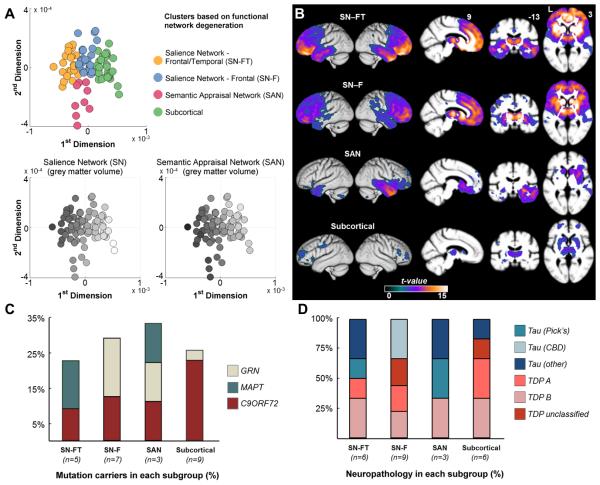
Design, setting and participants: In this retrospective observational study, 90 patients meeting the Frontotemporal Dementia Consortium consensus criteria for bvFTD underwent evaluation at the Memory and Aging Center of the Department of Neurology at University of California, San Francisco. Patients underwent a multidisciplinary clinical evaluation, including clinical demographics, genetic testing, symptom evaluation, neurologic examination, neuropsychological bedside testing, and socioemotional assessments. All patients underwent structural magnetic resonance imaging at their earliest evaluation at the memory clinic. From each patient's structural imaging scans, the mean volumes of 18 regions of interest (ROI) constituting the functional networks specifically vulnerable in bvFTD, including the salience network (SN), with key nodes in the frontoinsula and pregenual anterior cingulate, and the semantic appraisal network (SAN), anchored in the anterior temporal lobe and subgenual cingulate, were estimated. Principal component and cluster analyses of ROI volumes were used to identify patient clusters with anatomically distinct atrophy patterns. Data were collected from from June 19, 2002, to January 13, 2015.

Main outcomes and measures: Evaluation of brain morphology and other clinical features, including presenting symptoms, neurologic examination signs, neuropsychological performance, rate of dementia progression, and socioemotional function, in each patient cluster.

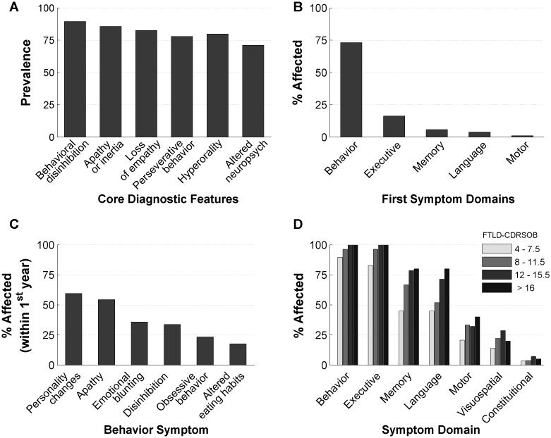
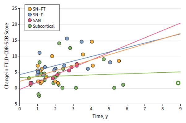
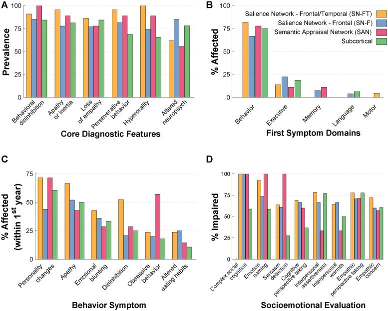
Results: Ninety patients (54 men [60%]; 36 women [40%]; mean [SD] age at evaluation, 55.1 [9.7] years) were included in the analysis. Four subgroups of patients with bvFTD with distinct anatomic patterns of network degeneration were identified, including 2 salience network-predominant subgroups (frontal/temporal [SN-FT] and frontal [SN-F]), a semantic appraisal network-predominant group (SAN), and a subcortical-predominant group. Subgroups demonstrated distinct patterns of cognitive, socioemotional, and motor symptoms, as well as genetic compositions and estimated rates of disease progression.

Conclusions and relevance: Divergent patterns of vulnerability in specific functional network components make an important contribution to the clinical heterogeneity of bvFTD. The data-driven anatomic classification identifies biologically meaningful anatomic phenotypes and provides a replicable approach to disambiguate the bvFTD syndrome.


Figures

Figure 1
Figure 1. Prevalence of FTDC criteria and major symptom domains in bvFTD
A: Prevalence of core diagnostic features of bvFTD patients. B: Percentage of patients with the initial symptoms in behavior, executive, memory, language, and motor domains. C: Rates of NACC behavioral symptom checklist categories in bvFTD patients during the first 12 months of presenting to memory clinic (n=94). Note that only the top six symptoms are shown in descending order from left to right. D: The rates of symptoms in each of the main domains, in bvFTD patients categorized according to their disease severity (i.e. FTLD-CDRSOB score). FTDC = Frontotemporal Dementia Criteria Consortium; FTLD-CDRSOB = Frontotemporal Lobar Degeneration modified Clinical Dementia Rating Sum of Boxes (CDRSOB); NACC = National Alzheimer Coordinating Center.
Figure 2
Figure 2. Anatomic subtypes of bvFTD
bvFTD patients are clustered into four distinct subgroups based on the degree of atrophy in 18 ROIs defining SN and SAN functional networks. The four clusters consist of two distinct SN affected groups – frontotemporal (SN-FT, the yellow dots) and frontal (SN-F, the blue dots), one SAN affected group (the pink dots), and one subcortical group (green dots). A (top panel): Each patient is colored according to their cluster assignment and plotted onto the first two dimensions of a principal component analysis based on the same 18 ROIs. A (bottom panels): The grey scale indicates the normalized volume of the sum of each patient’s ROIs representing the SN and SAN networks respectively, where darker colors show greater degree of volume loss. x and y axes are the same as for the top panel. B: Voxel based morphometry-derived atrophy maps of each bvFTD subgroup. T maps show the atrophy patterns compared to age matched normal controls (n = 44), and are superimposed onto whole-brain template derived from elderly normal controls. Effects are corrected for age, gender and total intracranial volume of each individual, and are corrected for family-wise error at the whole brain level at P<.05. C: Percentage of mutation carriers for C9ORF72, MAPT, or GRN mutations in each subgroup. D: Percentage of patients found to have specific classes of neuropathology associated with each bvFTD subgroup, among the subset of patients with autopsy diagnosis. Patients who were diagnosed as Tau-other pathology included two SN-FT patients with FTLD-tau with MAPT mutation, one SAN patient with FTDP17, and one subcortical patient with argyrophilic grain disease. CBD = corticobasal degeneration; C9ORF72 = chromosome 9 open reading frame 72 hexanucleotide expansions; FTDP-17=Frontotemporal dementia with parkinsonism-17; FTLD=Frontotemporal lobar degeneration; GRN = progranulin; MAPT microtubule-associated protein tau; ROI = region of interest; SAN = sematic appraisal network; SN = salience network; TDP = transactive response DNA-binding protein 43.
Figure 3
Figure 3. FTDC criteria, presenting symptoms and socioemotional dysfunction in bvFTD subgroups
A: Prevalence of core diagnostic features in each bvFTD subgroup. B: Percentage of patients in each bvFTD subgroup with the initial symptoms in behavior, executive, memory, language, and motor domains. C: Rates of NACC behavioral symptom checklist categories in each bvFTD subgroup during the first 12 months of presenting to memory clinic. Note that only the top six symptoms are shown in descending order from left to right. D: Rates of specific socioemotional impairments observed in each bvFTD subgroup. Socioemotional function was considered impaired at a Z-score<1.35 based on published normative samples of age-matched healthy controls. Complex social cognition, emotion naming and sarcasm detection (paralinguistic, i.e. voice prosody and facial expression) abilities were measured with The Awareness of Social Inference Test. Cognitive perspective taking was assessed with the UCSF Cognitive theory of Mind (cTOM) test. Interpersonal assertiveness and warmth were measured with the Dominance and Warmth subscales of the Interpersonal Adjective Scales. Empathy was measured with the Interpersonal Reactivity Index. FTDC = Frontotemporal Dementia Criteria Consortium; NACC = National Alzheimer Coordinating Center.

Comment in

References

    1. Neary D, Snowden JS, Gustafson L, et al. Frontotemporal lobar degeneration: a consensus on clinical diagnostic criteria. Neurology. 1998;51(6):1546–1554. - PubMed
    1. Snowden JS, Neary D, Mann DM. Frontotemporal dementia. Br J Psychiatry. 2002;180:140–143. - PubMed
    1. Hodges JR. Frontotemporal dementia (Pick's disease): clinical features and assessment. Neurology. 2001;56(11 Suppl 4):S6–10. - PubMed
    1. Pievani M, de Haan W, Wu T, Seeley WW, Frisoni GB. Functional network disruption in the degenerative dementias. Lancet Neurol. 2011;10(9):829–843. - PMC - PubMed
    1. Seeley WW, Crawford RK, Zhou J, Miller BL, Greicius MD. Neurodegenerative diseases target large-scale human brain networks. Neuron. 2009;62(1):42–52. - PMC - PubMed

Publication types

MeSH terms

LinkOut - more resources