Review
. 2016 Jun 30;13(7):540-55.
doi: 10.7150/ijms.15233. eCollection 2016.

Therapeutic Progress in Treating Vertebral Dissecting Aneurysms Involving the Posterior Inferior Cerebellar Artery

Affiliations
Review

Therapeutic Progress in Treating Vertebral Dissecting Aneurysms Involving the Posterior Inferior Cerebellar Artery

Lei Shi et al. Int J Med Sci. .

Abstract

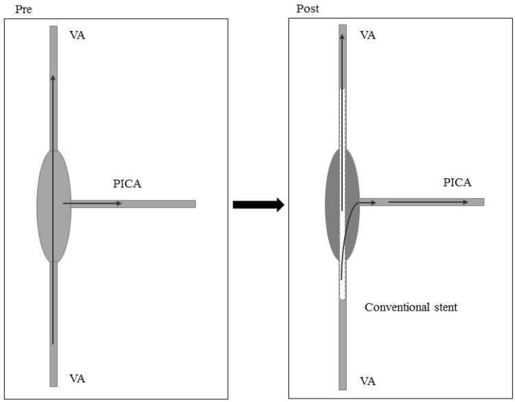
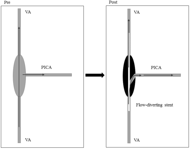
Among the variations of vertebral artery dissecting aneurysms (VDAs), VDAs involving the posterior inferior cerebellar artery (PICA), especially ruptured and high-risk unruptured aneurysms, are the most difficult to treat. Because the PICA is an important structure, serious symptoms may occur after its occlusion. Retained PICAs are prone to re-bleeding because VDAs are difficult to completely occlude. There is therefore confusion regarding the appropriate treatment for VDAs involving the PICA. Here, we used the PubMed database to review recent research concerning VDAs that involve the PICA, and we found that treatments for VDAs involving the PICA include (i) endovascular treatment involving the reconstruction of blood vessels and blood flow, (ii) occluding the aneurysm using an internal coil trapping or an assisted bypass, (iii) inducing reversed blood flow by occluding the proximal VDA or forming an assisted bypass, or (iv) the reconstruction of blood flow via a craniotomy. Although the above methods effectively treat VDAs involving the PICA, each method is associated with both a high degree of risk and specific advantages and disadvantages. The core problem when treating VDAs involving the PICA is to retain the PICA while occluding the aneurysm. Therefore, the method is generally selected on a case-by-case basis according to the characteristics of the aneurysm. In this study, we summarize the various current methods that are used to treat VDAs involving the PICA and provide schematic diagrams as our conclusion. Because there is no special field of research concerning VDAs involving the PICA, these cases are hidden within many multiple-cases studies. Therefore, this study does not review all relevant documents and may have some limitations. Thus, we have focused on the mainstream treatments for VDAs that involve the PICA.

Keywords: Vertebral artery dissecting aneurysms; posterior inferior cerebellar artery; therapeutic progress..


Conflict of interest statement

Competing Interests: The authors have declared that no competing interest exists.

Figures

Figure 1
Figure 1
Conventional stent implantation for vascular reconstruction. Pre: preoperation, Post: postoperation, VA: vertebral artery, PICA: posterior inferior cerebral artery.
Figure 2
Figure 2
Stent-assisted coil occlusion for vascular reconstruction. Pre: preoperation, Post: postoperation, VA: vertebral artery, PICA: posterior inferior cerebral artery.
Figure 3
Figure 3
Blood flow-diverting stent for vascular reconstruction. Pre: preoperation, Post: postoperation, VA: vertebral artery, PICA: posterior inferior cerebral artery.
Figure 4
Figure 4
Internal coil trapping of aneurysms involving the sacrificed of the PICA. Pre: preoperation, Post: postoperation, VA: vertebral artery, PICA: posterior inferior cerebral artery.
Figure 5
Figure 5
A: OA-PICA bypass-assisted internal coil trapping of an aneurysm in which the PICA is sacrificed; B: PICA-PICA bypass after internal coil trapping of the aneurysm with PICA sacrifice. Pre: preoperation, Post: postoperation, VA: vertebral artery, PICA: posterior inferior cerebral artery, OA: occipital artery.
Figure 6
Figure 6
Internal coil trapping for aneurysm occlusion with stent implantation from the VA to the PICA. Pre: preoperation, Post: postoperation, VA: vertebral artery, PICA: posterior inferior cerebral artery.
Figure 7
Figure 7
A: Proximal endovascular occlusion of the VDA; B: Proximal surgical clipping of the VDA. Pre: preoperation, Post: postoperation, VA: vertebral artery, PICA: posterior inferior cerebral artery.
Figure 8
Figure 8
Proximal occlusion or clipping of the VDA or bypass assistance. Pre: preoperation, Post: postoperation, VA: vertebral artery, PICA: posterior inferior cerebral artery.
Figure 9
Figure 9
PICA reimplantation to the VA after VDA trapping. Pre: preoperation, Post: postoperation, VA: vertebral artery, PICA: posterior inferior cerebral artery.
Figure 10
Figure 10
PICA-PICA bypass after VDA trapping. Pre: preoperation, Post: postoperation, VA: vertebral artery, PICA: posterior inferior cerebral artery.
Figure 11
Figure 11
Reconstruction of the PICA with a vascular graft with the graft end on the PICA side after VDA trapping. Pre: preoperation, Post: postoperation, VA: vertebral artery, PICA: posterior inferior cerebral artery
Figure 12
Figure 12
Reconstruction of the PICA with a vascular graft, with the graft end at the PICA end after VDA trapping. Pre: preoperation, Post: postoperation, VA: vertebral artery, PICA: posterior inferior cerebral artery.
Figure 13
Figure 13
Reconstruction of the VA and PICA after VDA trapping. Pre: preoperation, Post: postoperation, VA: vertebral artery, PICA: posterior inferior cerebral artery.
Figure 14
Figure 14
Reconstruction of the VA and PICA after VDA removal. Pre: preoperation, Post: postoperation, VA: vertebral artery, PICA: posterior inferior cerebral artery.
Figure 15
Figure 15
Figures showing typical cases. A: CT revealing an intracranial subarachnoid hemorrhage concentrated in the suprasellar cistern and posterior ambient cistern. B-C: CTA and DSA revealing a left VDA in which the PICA originates from the aneurysm. D: Angiography of contralateral vertebral artery revealing the reversal of blood flow to the aneurysm and shadows showing the PICA. E-F: Coils were filled in the weak points of the aneurysm, covering LVIS stents, and the distal VA of the aneurysm developed vasospasm. G: After one week, DSA revealed that the vasospasm was released. H: The occlusion of the VA in the extracranial vertebral artery (segment V2). Angiography of the contralateral vertebral artery revealed the reversal of blood flow to the aneurysm and clear images of the PICA.

References

    1. Hernandez-Duran S, Ogilvy CS. Clinical outcomes of patients with vertebral artery dissection treated endovascularly: a meta-analysis. Neurosurg Rev. 2014;37:569–77. - PubMed
    1. Iihara K, Sakai N, Murao K, Sakai H, Higashi T, Kogure S. et al. Dissecting aneurysms of the vertebral artery: a management strategy. J Neurosurg. 2002;97:259–67. - PubMed
    1. Fine AD, Cardoso A, Rhoton AL Jr. Microsurgical anatomy of the extracranial-extradural origin of the posterior inferior cerebellar artery. J Neurosurg. 1999;91:645–52. - PubMed
    1. Matsukawa H, Shinoda M, Fujii M, Takahashi O, Uemura A, Niimi Y. Basilar extension and posterior inferior cerebellar artery involvement as risk factors for progression of the unruptured spontaneous intradural vertebral artery dissection. J Neurol Neurosurg Psychiatry. 2014;85:1049–54. - PubMed
    1. Sano H, Kato Y, Okuma I, Yamaguchi S, Ninomiya T, Arunkumar R. et al. Classification and treatment of vertebral dissecting aneurysm. Surg Neurol. 1997;48:598–605. - PubMed

MeSH terms