

Saphenous vein graft aneurysm: A case report
- PMID: 27429630
- PMCID: PMC4933749
Saphenous vein graft aneurysm: A case report
Abstract
Background: Saphenous vein graft aneurysms (SVGAs) are rare seen issues after coronary artery bypass graft (CABG) operation which may lead to major complications including compression of adjacent structure, myocardial ischemia, rupture, and even death.
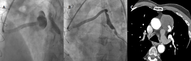
Case report: We report a patient with recurrent SVGA and its treatment by percutaneous intervention with a covered stent, the diagnostic and treatment procedure were based on contrast enhanced computed tomography and myocardial perfusion scintigraphy (MPS).
Conclusion: Multimodality imaging is required to demonstrate the true size and complications of the SVGA, the relationship among the adjacent structure, and to assess ischemia and size of myocardial territory supplied by the aneurysmal graft to decide treatment strategy.
Keywords: Computed Tomography; Coronary Aneurysm; Coronary Artery Bypass Grafting; Saphenous Vein; Stents.
Figures


References
-
- Ramirez FD, Hibbert B, Simard T, Pourdjabbar A, Wilson KR, Hibbert R, et al. Natural history and management of aortocoronary saphenous vein graft aneurysms: a systematic review of published cases. Circulation. 2012;126(18):2248–56. - PubMed
-
- Douglas BP, Bulkley BH, Hutchins GM. Infected saphenous vein coronary artery bypass graft with mycotic aneurysm. Fatal dehiscence of the proximal anastomosis. Chest. 1979;75(1):76–7. - PubMed
-
- Ennis BM, Zientek DM, Ruggie NT, Billhardt RA, Klein LW. Characterization of a saphenous vein graft aneurysm by intravascular ultrasound and computerized three-dimensional reconstruction. Cathet Cardiovasc Diagn. 1993;28(4):328–31. - PubMed
-
- Werthman PE, Sutter FP, Flicker S, Goldman SM. Spontaneous, late rupture of an aortocoronary saphenous vein graft. Ann Thorac Surg. 1991;51(4):664–6. - PubMed
Publication types
LinkOut - more resources
Full Text Sources