. 2016 Feb;3(2):1500223.
doi: 10.1002/advs.201500223. Epub 2015 Oct 27.

Biodegradable and Renal Clearable Inorganic Nanoparticles

Affiliations

Biodegradable and Renal Clearable Inorganic Nanoparticles

Emily B Ehlerding et al. Adv Sci (Weinh). 2016 Feb.

Abstract

Personalized treatment plans for cancer therapy have been at the forefront of oncology research for many years. With the advent of many novel nanoplatforms, this goal is closer to realization today than ever before. Inorganic nanoparticles hold immense potential in the field of nano-oncology, but have considerable toxicity concerns that have limited their translation to date. In this review, an overview of emerging biologically safe inorganic nanoplatforms is provided, along with considerations of the challenges that need to be overcome for cancer theranostics with inorganic nanoparticles to become a reality. The clinical and preclinical studies of both biodegradable and renal clearable inorganic nanoparticles are discussed, along with their implications.


Figures

Scheme 1
Scheme 1
Schematic illustrations of biodegradable (a–c) and renal clearable inorganic nanoparticles (d–f). a) Ultra‐small superparamagnetic iron oxide (USPIO) nanoparticle for magnetic resonance imaging. b) Biodegradable biopolymer coated luminescent porous silicon nanoparticles (LPSiNP). c) Functionalized hollow mesoporous silica nanoparticle (HMSN) for positron emission tomography imaging, optical imaging, and tumor targeted drug delivery. d) Renal clearable cysteine coated quantum dot (QD). e) Ultra‐small fluorescent silica nanoparticle (also known as C dot) functionalized with targeting ligands (cRGDY), poly(ethylene glycol) and radioisotope (124I) for tumor targeted multimodality imaging. f) Glutathione (GS), or poly(ethylene glycol) (PEG) coated 2–3 nm sized AuNPs.
Figure 1
Figure 1
a) Schematic illustration of in vivo degradation process of the biopolymer‐coated LPSiNPs. b) Degradation and change of photoluminescence when incubating LPSiNPs in PBS at 37 °C. c) In vivo biodistribution and biodegradation of LPSiNPs over a period of 4 weeks. d) In vivo image showing the clearance of LPSiNPs into the bladder at 1 h post injection. Li (liver) and Bl (bladder). Reproduced with permission.9 Copyright 2009, Nature Publishing Group.
Figure 2
Figure 2
TEM images showing the degradation of MSN (pore size: ≈10 nm) in simulated body fluid (SBF) at 37 °C. a) 0 h, b) 4 h, c) 12 h and d) 24 h after soaking in SBF. Scale bar: 100 nm. Reproduced with permission.40 Copyright 2014, American Chemical Society.
Figure 3
Figure 3
a) Schematic illustration of ultra‐small QDs. b) Renal clearable cutoff study. Top: color photos of un‐injected bladders. Bottom: fluorescence images of un‐injected bladders. Middle: fluorescence images at 4 h post injection of different QDs (hydrodynamic size range: 4–8 nm). Reproduced with permission.10 Copyright 2007, Nature Publishing Group. c) A schematic illustration of ultra‐small QDs functionalized with targeting ligands and NIR) fluorophores. d) In vivo NIR fluorescence imaging of GPI (a ligand which targets to prostate‐specific membrane antigen [PSMA]) and 800‐CW functionalized QDs (or QD‐CW‐GPI) in PSMA positive (LNCaP) and negative (PC‐3) tumor models. Reproduced with permission.11 Copyright 2010, Nature Publishing Group.
Figure 4
Figure 4
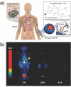
a) Schematic illustration of the use of cRGDY peptide functionalized, 124 I‐labeled C dot as hybrid (PET‐optical) imaging nanoparticle (124I‐cRGDY–PEG–C dots) in a human patient. b) Maximum intensity projection PET images at 3, 24, and 72 h after intravenous injection of 124I‐cRGDY–PEG–C dots revealed probe activity in bladder (*), heart (yellow arrow), and bowel (white arrowhead). Reproduced with permission.5 Copyright 2014, American Association for the Advancement of Science.
Figure 5
Figure 5
a) TEM image of GS‐[198Au]AuNP. Scale bare: 5 nm. b)ynamic light scattering analysis of GS‐[198Au]AuNP showing a HD size of 3.0 ± 0.4 nm. Core size: 2.6 ± 0.3 nm (inset). c) In vivo SPECT (left) and fluorescence images (right) of balb/c mice after tail vein injection of GS‐[198Au]AuNP 1 h post injection. Reproduced with permission.50 d) TEM image of PEG1k‐AuNP. e) Dynamic light scattering analysis of PEG1k‐AuNP showing a HD size of 5.5 ± 0.4 nm. Core size: 2.3 ± 0.3 nm (inset). f) In vivo NIR fluorescence imaging of PEG1k‐AuNP in MCF‐7 tumor‐bearing nude mice after tail veil injection at 5 and 48 h (EPR based passive targeting). Reproduced with permission.48

References

    1. Siegel R., Ma J., Zou Z., Jemal A., Ca‐Cancer J. Clin. 2014, 64, 9. - PubMed
    1. Wistuba I. I., Gelovani J. G., Jacoby J. J., Davis S. E., Herbst R. S., Nat. Rev. Clin. Oncol. 2011, 8, 135. - PubMed
    1. Thakor A. S., Gambhir S. S., Ca‐Cancer J. Clin. 2013, 63, 395. - PubMed
    1. Benezra M., Penate‐Medina O., Zanzonico P. B., Schaer D., Ow H., Burns A., DeStanchina E., Longo V., Herz E., Iyer S., Wolchok J., Larson S. M., Wiesner U., Bradbury M. S., J. Clin. Invest. 2011, 121, 2768. - PMC - PubMed
    1. Phillips E., Penate‐Medina O., Zanzonico P. B., Carvajal R. D., Mohan P., Ye Y., Humm J., Gönen M., Kalaigian H., Schöder H., Strauss H. W., Larson S. M., Wiesner U., Bradbury M. S., Sci.Transl. Med. 2014, 6, 260ra149. - PMC - PubMed