

Minor Antigen Vaccine-Sensitized DLI: In Vitro Responses Do Not Predict In Vivo Effects
- PMID: 27430015
- PMCID: PMC4943762
- DOI: 10.1097/TXD.0000000000000583
Minor Antigen Vaccine-Sensitized DLI: In Vitro Responses Do Not Predict In Vivo Effects
Abstract
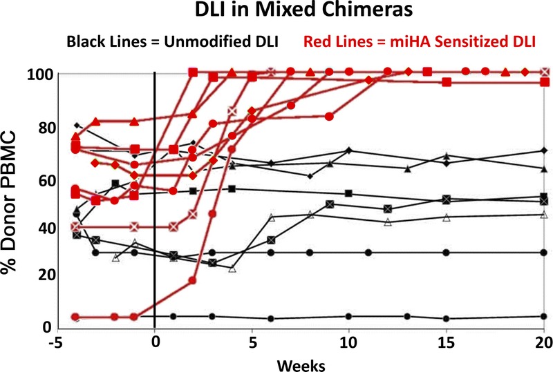
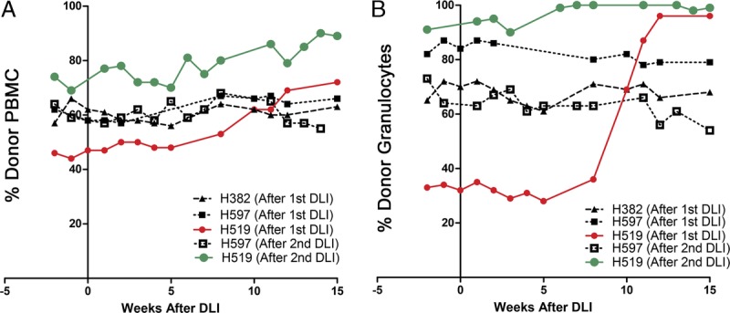
We reported on a pilot study of minor histocompatibility antigen vaccination using constructs expressing male-specific gene disparities of selected mouse CDNA on Y and sex determining region Y in the canine model. We performed reduced-intensity hematopoietic cell transplantation with female donors and male recipients, producing stable mixed donor-recipient hematopoietic chimeras. We then performed a vaccine series in three female transplant donors followed by donor lymphocyte infusion (DLI) into their respective mixed chimeras. One mixed chimera experienced a significant shift in the percentage of donor chimerism, but no response occurred in the other 2 recipients. We then hypothesized that inadequate donor sensitization was responsible for these results.
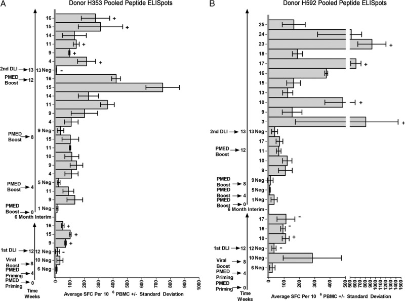
Methods: To test this hypothesis, we added 4 monthly booster vaccinations to 2 of the original hematopoietic cell transplantation donors, including the donor that drove the partial response, followed by a second DLI.
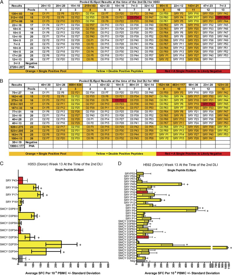
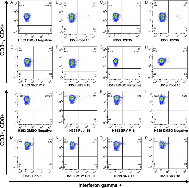
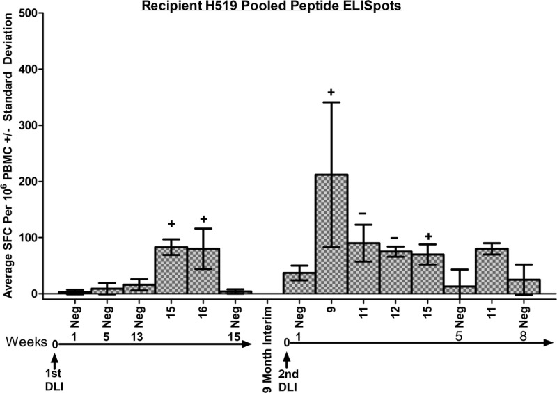
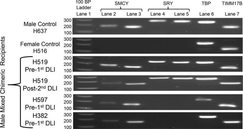
Results: Strong T cell responses were shown by ELISpot and confirmed by intracellular cytokine staining in both donors. A second DLI resulted in a further increase in donor chimerism in the same mixed chimera that experienced the previous increase, but no change in donor chimerism was again seen in the other recipient. Evaluation of RNA expression of the target antigens demonstrated that conversion occurred in the recipient that expressed both selected mouse CDNA on Y and sex determining region Y.
Conclusions: T cell responses against Y chromosome-encoded disparities were not necessarily sufficient to drive in vivo female antimale responses. Other factors including the presence of specific haplotypes or the heterogeneous expression of the target antigen may affect T cell responses against minor histocompatibility antigens. These results warrant future vaccine studies in a larger transplant cohort using epigenetic modulation of the recipient to promote target gene expression.
Conflict of interest statement
The authors are grateful for research funding from the National Institutes of Health, Bethesda, MD, grants P01 CA078902, P30 CA015704, K12 CA76930, P30 DK56465, support from Gabrielle's Angel Foundation for Cancer Research (SLR), and by awards from the Joseph Steiner Krebsstiftung, Bern, Switzerland and Lupin Foundation, Metairie, LA (RS).
The authors declare no conflicts of interest.
S.L.R., B.S., and S.S.G. participated in research design, performance of research, data analysis, and writing of the article. D.H.F. and J.T.F. contributed new reagents, performance of research, and participated in research design. R.S. participated in research design and writing of the article.
Figures
Similar articles
-
Adoptive immunotherapy against kidney targets in dog-leukocyte antigen-identical mixed hematopoietic canine chimeras.Transplantation. 2003 Feb 15;75(3):268-74. doi: 10.1097/01.TP.0000045224.52516.FC. Transplantation. 2003. PMID: 12589144
-
IL-2 does not enhance the conversion to complete donor chimerism following nonmyeloablative hematopoietic cell transplantation in dogs.Bone Marrow Transplant. 2003 Jun;31(11):1027-31. doi: 10.1038/sj.bmt.1704046. Bone Marrow Transplant. 2003. PMID: 12774055
-
Donor lymphocyte infusion-mediated graft-versus-leukemia effects in mixed chimeras established with a nonmyeloablative conditioning regimen: extinction of graft-versus-leukemia effects after conversion to full donor chimerism.Transplantation. 2003 Jul 27;76(2):297-305. doi: 10.1097/01.TP.0000072014.83469.2D. Transplantation. 2003. PMID: 12883182
-
Early host CD8 T-cell recovery and sensitized anti-donor interleukin-2-producing and cytotoxic T-cell responses associated with marrow graft rejection following nonmyeloablative allogeneic bone marrow transplantation.Exp Hematol. 2003 Jul;31(7):609-21. doi: 10.1016/s0301-472x(03)00082-1. Exp Hematol. 2003. PMID: 12842706
-
Alloreactivity as therapeutic principle in the treatment of hematologic malignancies. Studies of clinical and immunologic aspects of allogeneic hematopoietic cell transplantation with nonmyeloablative conditioning.Dan Med Bull. 2007 May;54(2):112-39. Dan Med Bull. 2007. PMID: 17521527 Review.
References
-
- Goulmy E. Minor histocompatibility antigens: allo target molecules for tumor-specific immunotherapy (Review). Cancer J. 2004; 10: 1– 7. - PubMed
-
- Spierings E. Minor histocompatibility antigens: past, present, and future (Review). Tissue Antigens. 2014; 84: 374– 60. - PubMed
-
- Storb R, Yu C, Wagner JL, et al. Stable mixed hematopoietic chimerism in DLA-identical littermate dogs given sublethal total body irradiation before and pharmacological immunosuppression after marrow transplantation. Blood. 1997; 89: 3048– 3054. - PubMed
-
- Georges GE, Storb R, Thompson JD, et al. Adoptive immunotherapy in canine mixed chimeras after nonmyeloablative hematopoietic cell transplantation. Blood. 2000; 95: 3262– 3269. - PubMed
Grants and funding
LinkOut - more resources
Full Text Sources
Other Literature Sources
