. 2016 Jun 16;1(9):e86177.
doi: 10.1172/jci.insight.86177.

A flexible, multilayered protein scaffold maintains the slit in between glomerular podocytes

Affiliations

A flexible, multilayered protein scaffold maintains the slit in between glomerular podocytes

Florian Grahammer et al. JCI Insight. .

Abstract

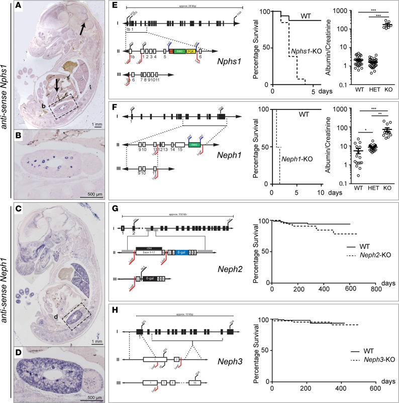
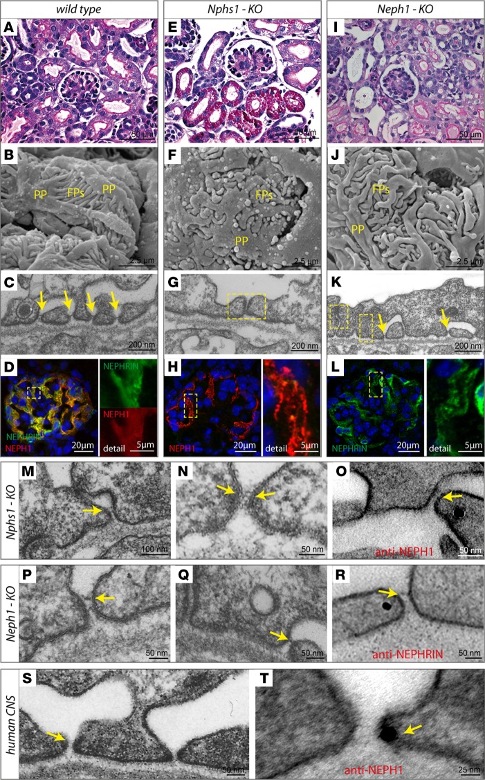
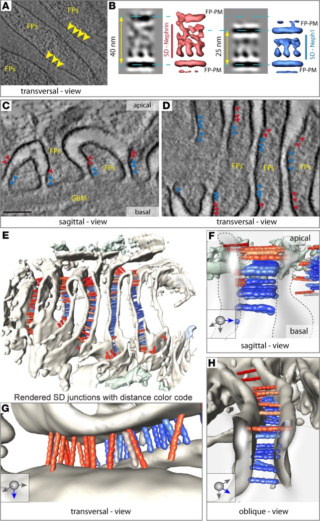
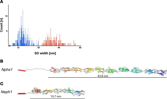
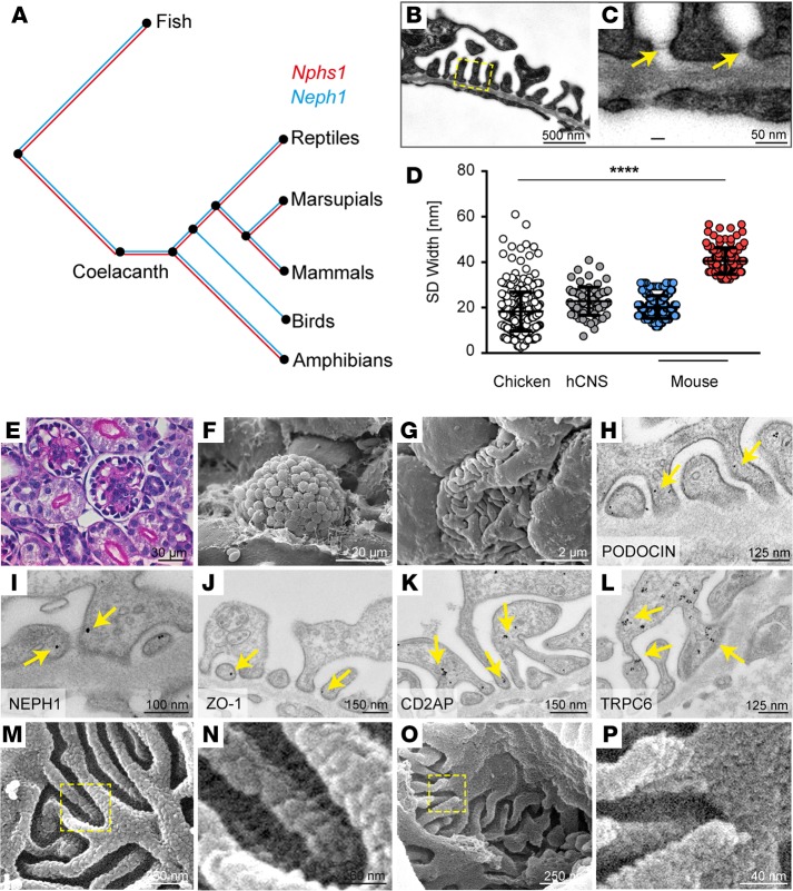
Vertebrate life critically depends on renal filtration and excretion of low molecular weight waste products. This process is controlled by a specialized cell-cell contact between podocyte foot processes: the slit diaphragm (SD). Using a comprehensive set of targeted KO mice of key SD molecules, we provided genetic, functional, and high-resolution ultrastructural data highlighting a concept of a flexible, dynamic, and multilayered architecture of the SD. Our data indicate that the mammalian SD is composed of NEPHRIN and NEPH1 molecules, while NEPH2 and NEPH3 do not participate in podocyte intercellular junction formation. Unexpectedly, homo- and heteromeric NEPHRIN/NEPH1 complexes are rarely observed. Instead, single NEPH1 molecules appear to form the lower part of the junction close to the glomerular basement membrane with a width of 23 nm, while single NEPHRIN molecules form an adjacent junction more apically with a width of 45 nm. In both cases, the molecules are quasiperiodically spaced 7 nm apart. These structural findings, in combination with the flexibility inherent to the repetitive Ig folds of NEPHRIN and NEPH1, indicate that the SD likely represents a highly dynamic cell-cell contact that forms an adjustable, nonclogging barrier within the renal filtration apparatus.


Figures

Figure 1
Figure 1. Lack of NEPHRIN or NEPH1, but not NEPH2 or NEPH3, causes early lethality and severe nephrotic kidney disease.
(A and B) At E13.5, Nphs1 showed a restricted expression pattern in renal glomeruli, pancreas, and cerebellum anlage. First arrow, cerebellum anlage; second arrow, pancreas; box labeled b is the magnified area shown in B. (C and D) At E13.5, Neph1 was broadly expressed in the central nervous system, lung, kidney, and gut. Box labeled d is the magnified area shown in D. (EH) Targeting strategy and survival analysis of constitutive Nphs1, Neph1, Neph2, and Neph3 KO mice. Constitutive KO of Nphs1 or Neph1 led to early perinatal lethality, whereas mice deficient in Neph2 or Neph3 did not die prematurely (log rank [Mantel-Cox] test was used with at least 10 animals per genotype; P < 0.01 for Nphs1–/– and P < 0.0001 for Neph1–/–; no difference for Neph2–/– and Neph3–/–). Both Nphs1–/– and Neph1–/– mice presented with massive proteinuria (at least 5 animals per genotype and allele were analyzed for urinary albumin and creatinine; *P < 0.05, **P < 0.01, ***P < 0.001).
Figure 2
Figure 2. NEPHRIN and NEPH1 can form rudimentary SDs independently of each other.
(AC and EG) In contrast to control mice, Nphs1–/– mice had protein-filled proximal tubular cells, broadened primary (PP) and shortened secondary foot processes (FP), and what appeared to be tight junctional cell-cell contacts between effaced foot processes. Yellow arrows represent SDs slit diaphragms; yellow squares represent tight junctions. (IK) Neph1–/– mice appeared to have misdirected foot processes but, at least at early stages of glomerular maturation, were able to form SD-like junctions. (D, H, and L) Immunoreactivity for NEPH1 was largely maintained in Nphs1–/–, as was staining intensity for NEPHRIN in Neph1–/–. (MO) Further evaluation of Nphs1-deficient glomeruli demonstrated thin and short strand-like junctions between incompletely effaced foot processes that showed NEPH1 immunoreactivity. (PR) Similarly, in Neph1–/– mice, we detected SD-like junctions that often appeared dislocated apically and had NEPHRIN immunoreactivity. (S and T) In tissue from human CNS patients with remaining open slits, SD-like junctions could be visualized, showing immunoreactivity for NEPH1.
Figure 3
Figure 3. The SD is a multilayered, bipartite protein scaffold.
(A) CET (cryo-electrontomography) of vitreous sections: Tangential computational 2-nm thick section through the base of several foot processes (FP). Individual densities (indicated by the yellow arrowheads) can be seen spanning the area between 2 FP. They are quasiperiodically arranged and have similar thickness, without a dense midline. (B) Subtomogram averaging of these densities reveals 2 predominant classes; the first class has a intermembrane distance of 40 nm (corresponding isosurface representation shown in red). The second has an intermembrane distance of 25 nm (isosurface representation shown in blue). For both entities, quasiperiodic strands with a thickness of 3 nm can be seen spanning the membranes. With increasing distance, the strands appear more disordered, while the shorter ones are rather regularly arranged. A dense midline cannot be discerned. (C) Computational sagittal section (10-nm thick) from a PET tomogram showing the typical appearance of podocytes. Several strands cross the individual FPs with the narrower end of the slit toward the GBM, while the broader opening faces Bowman’s space. The colored arrowheads point to individual strands. Red arrowheads correspond to longer strands, while blue arrowheads mark shorter strands. (D) Transversal computational section (10-nm thick) (in a similar orientation as A) showing the quasiperiodic arrangement of several strands (color code as in C). (E) Surface visualization (3D) seen from Bowman’s capsule showing several FP and hundreds of bridging strands color-coded depending on their length (red 40 nm, blue 25 nm; color variation indicating deviations from these). The histogram of their length variation is shown in Figure 4A. (F) Typical sagittal view of 2 FP, showing 3 layers of 25-nm strands, which we attribute to NEPH1 (blue, and 5 Ig repeats) and one layer of 40-nm strands that we attribute to NEPHRIN (red, and 9 Ig repeats). (G) Transversal view showing a quasiperiodic arrangement of NEPHRIN molecules. (H) Oblique view from Bowman’s capsule onto the molecular arrangement of NEPHRIN and NEPH1.
Figure 4
Figure 4. Structural modeling of NPHS1 and NEPH1 based on their homology to TITIN.
(A) Length distribution of individual strands within mouse foot processes in PET reconstructions gives 2 gaussian-like distributions centered at 45 nm and 23 nm. (B) For mouse NPHS1, databases (uniprot: Q9QZS7, ncbi: NP_062332.2) predict 8 N-terminal immunoglobulin type C2 domains and 1 C-terminal fibronectin type III domain. A ninth Ig domain (between Ig6 and Ig7, aa 650–753) was predicted with sequence alignments of all mouse NPHS1 Ig domains and with the use of PHYRE2 (36). Straight length for NEPHRIN was estimated at 43.8 nm. (C) In accordance with the literature, mouse NEPH1 (uniprot Q80W68, ncbi: NM_019459.2) consists of 5 N-terminal immunoglobulin type C2 domains. In the case of NEPH1, straight length was estimated to be 19.7 nm.
Figure 5
Figure 5. Evolutionary concept of a NEPH1-based SD formation in birds.
(A) Phylogenetic analysis of Nphs1 and Neph1 throughout the animal kingdom. (B and C) Coelacanths contain both genes, whereas Nphs1 was lost only in birds. TEM reveals that foot processes are evenly spaced, and multiple layers of slit diaphragms can be detected in Gallus gallus. (D) Analysis of SD width illustrates that the chicken SD is similar to the one found in human CNS (hCNS), while mice have an SD containing both narrow and wide cell-cell junctions (chicken, n = 391; human, n = 77; mouse NEPH1, n = 360; NEPHRIN, n = 77; all n quantities refer to individual measurements in at least n = 3 different biological samples). ****P < 0.0001 by ANOVA. (EG) Light microscopy images demonstrate that podocyte cell bodies in G. gallus are found more toward the edge of the glomerular field directly opposite Bowman’s capsule, whereas SEM reveals broadened primary and shortened secondary foot processes compared with mice. (HL) Established SD molecules — including PODOCIN, NEPH1, ZO-1, CD2AP, and TRPC6 — can be detected at the chicken SD. Arrows indicate immunogold particles. (M and N) High-resolution SEM of mouse glomeruli reveals individual strands of molecules crossing the slit above an interwoven molecular mesh situated in the lower part of this intercellular junction. (O and P) Chicken glomeruli contain the mesh but lack the individual strands. Boxes in M and O indicate magnified area shown in N and P, respectively.

Comment in

Similar articles

Cited by

References

    1. Kestila M, et al. Positionally cloned gene for a novel glomerular protein--nephrin--is mutated in congenital nephrotic syndrome. Molecular Cell. 1998;1(4):575–582. doi: 10.1016/S1097-2765(00)80057-X. - DOI - PubMed
    1. Sellin L, Huber TB, Gerke P, Quack I, Pavenstädt H, Walz G. NEPH1 defines a novel family of podocin interacting proteins. FASEB J. 2003;17(1):115–117. - PubMed
    1. Donoviel DB, et al. Proteinuria and perinatal lethality in mice lacking NEPH1, a novel protein with homology to NEPHRIN. Mol Cell Biol. 2001;21(14):4829–4836. doi: 10.1128/MCB.21.14.4829-4836.2001. - DOI - PMC - PubMed
    1. Gerke P, et al. NEPH2 is located at the glomerular slit diaphragm, interacts with nephrin and is cleaved from podocytes by metalloproteinases. J Am Soc Nephrol. 2005;16(6):1693–1702. doi: 10.1681/ASN.2004060439. - DOI - PubMed
    1. Ihalmo P, Palmén T, Ahola H, Valtonen E, Holthöfer H. Filtrin is a novel member of nephrin-like proteins. Biochem Biophys Res Commun. 2003;300(2):364–370. doi: 10.1016/S0006-291X(02)02854-1. - DOI - PubMed