. 2016 Sep 2;119(6):e91-e103.
doi: 10.1161/CIRCRESAHA.116.308955. Epub 2016 Jul 18.

LNK/SH2B3 Loss of Function Promotes Atherosclerosis and Thrombosis

Affiliations

LNK/SH2B3 Loss of Function Promotes Atherosclerosis and Thrombosis

Wei Wang et al. Circ Res. .

Abstract

Rationale: Human genome-wide association studies have revealed novel genetic loci that are associated with coronary heart disease. One such locus resides in LNK/SH2B3, which in mice is expressed in hematopoietic cells and suppresses thrombopoietin signaling via its receptor myeloproliferative leukemia virus oncogene. However, the mechanisms underlying the association of LNK single-nucleotide polymorphisms with coronary heart disease are poorly understood.

Objective: To understand the functional effects of LNK single-nucleotide polymorphisms and explore the mechanisms whereby LNK loss of function impacts atherosclerosis and thrombosis.

Methods and results: Using human cord blood, we show that the common TT risk genotype (R262W) of LNK is associated with expansion of hematopoietic stem cells and enhanced megakaryopoiesis, demonstrating reduced LNK function and increased myeloproliferative leukemia virus oncogene signaling. In mice, hematopoietic Lnk deficiency leads to accelerated arterial thrombosis and atherosclerosis, but only in the setting of hypercholesterolemia. Hypercholesterolemia acts synergistically with LNK deficiency to increase interleukin 3/granulocyte-macrophage colony-stimulating factor receptor signaling in bone marrow myeloid progenitors, whereas in platelets cholesterol loading combines with Lnk deficiency to increase activation. Platelet LNK deficiency increases myeloproliferative leukemia virus oncogene signaling and AKT activation, whereas cholesterol loading decreases SHIP-1 phosphorylation, acting convergently to increase AKT and platelet activation. Together with increased myelopoiesis, platelet activation promotes prothrombotic and proatherogenic platelet/leukocyte aggregate formation.

Conclusions: LNK (R262W) is a loss-of-function variant that promotes thrombopoietin/myeloproliferative leukemia virus oncogene signaling and platelet and leukocyte production. In mice, LNK deficiency is associated with both increased platelet production and activation. Hypercholesterolemia acts in platelets and hematopoietic progenitors to exacerbate thrombosis and atherosclerosis associated with LNK deficiency.

Keywords: atherosclerosis; cholesterol; hematopoiesis; hypercholesterolemia; thrombosis.


Figures

Figure 1
Figure 1. Association of the LNK TT risk SNP with HSC expansion and increased megakaryopoiesis
Flow cytometry overview of HSCs (LinCD34+CD38loCD90+CD45RA) (a) and HSC percentage in CD34+ (b) or LinCD34+CD38lo (c) cord blood cell fractions. p-STAT5 (d) and p-ERK1/2 (e) levels in CD34+ cells. Megakaryocyte (Mk) colony number (f,g) and Mk counts per colony (h). Normalized Lnk mRNA levels in CD34+ cells (i). j) Allele-specific LNK expression of human cord blood CD34+ cells.
Figure 2
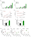
Figure 2. Hypercholesterolemia markedly increases myelopoiesis in Lnk−/− BM recipient
Hematopoietic stem and progenitor cells in BM (a), peripheral neutrophil, monocyte and platelet counts (b), cell surface CBS levels (c) and platelet neutrophil/monocyte aggregates (e). n=14–15. (d) p-Erk and p-Stat levels in BM cells with and without stimulation of IL-3 (100ng/ml) and GM-SCF (40ng/ml). Black bars represent chow feeding and green bars represent WTD feeding for 12 weeks. *, **, *** denote p<0.05, <0.01 and <0.001 for WTD-fed Lnk −/− vs WT or chow-fed Lnk −/− vs WT. ^, ^^, ^^^ denote p<0.05, <0.01 and <0.001 for chow-fed Lnk −/− vs WTD-fed Lnk −/− or chow-fed WT vs WTD-fed WT.
Figure 3
Figure 3. Hypercholesterolemia markedly increases platelet reactivity in Lnk−/− BM recipient
Surface P-selectin (a) and active integrin αIIbβ3 (JON/A) (b) levels on washed platelets with or without PAR4 agonist (AYPGKF, 100 μM) stimulation (n=5). Surface P-selectin (c) and active integrin αIIbβ3 (JON/A) (d) levels on platelets in whole blood with or without AYPGKF (100 μM) stimulation (n=5). Washed platelet aggregation upon AYPGKF (100μM) (e) or ADP (20 μM) (f) stimulation (n=4–5). TPO receptor (Mpl) levels on platelet surface (g), platelet TPO internalization (h) and plasma TPO levels (i) in WT or Lnk−/− BM recipient mice. (n=3–7) For 3a–3f, black bars represent chow feeding and green bars represent WTD feeding for 12 weeks. *, **, *** denote p<0.05, <0.01 and <0.001 for WTD-fed Lnk −/− vs WT or chow-fed Lnk −/− vs WT. ^, ^^, ^^^ denote p<0.05, <0.01 and <0.001 for chow-fed Lnk −/− vs WTD-fed Lnk −/− or chow-fed WT vs WTD-fed WT.
Figure 4
Figure 4. Cholesterol loading increases while cholesterol unloading decreases platelet and PKC activity
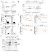
(a) Platelets from the chow-fed recipients were loaded with or without cholesterol and then stimulated with AYPGKF (100μM). Surface P-selectin levels were shown (n=4). (b) Platelets from the chow-fed recipients were loaded with or without cholesterol and then stimulated with or without AYPGKF (50μM). PKC activity was estimated (n=3). AYPGKF-induced surface P-selectin exposure (c) or aggregation (d) of platelets from the WTD-fed recipients, with or without rHDL (50 μg/ml) treatment. (e, f) similar as in (c) or (d) except that cyclodextrin (CD) (3mM) replaced HDL for the treatment (n=4). (g) PKC activity in platelets from WTD-fed recipients was estimated, with and without AYPGKF (100μM) and HDL (50μg/ml) treatment (n=3). For 3c and 3e, * p<0.05 between indicated groups; # p<0.05 between control (WTD) and CD or HDL treatment groups.
Figure 5
Figure 5. LNK loss of function and cholesterol enrichment increase platelet AKT activation. (a)
TPO/Mpl signaling in resting platelets of WT and Lnk −/− BM recipient mice without TPO treatment. (b) AKT activity in platelets from WT and Lnk −/− BM recipient mice with and without platelet agonist AYPGKF stimulation. (c) effect of ex ex vivo cholesterol loading on AKT activity from platelets of chow fed mice. (d) effect of Jak2 and AKT inhibitors on platelets of WT and Lnk −/− BM recipient mice (n=4). (e) p-SHIP1 level in platelets of WT and Lnk −/− BM recipient mice with and without AYPGKF stimulation. (f) change of AKT activity after HDL and Lyn tyrosine kinase activator Tolimidone treatment in Lnk−/− platelets from WTD fed mice. (g) p-SHIP1 levels in WT and Lynkd/kd platelets upon AYPGKF stimulation. (h) platelet activity of Lynkd/kd mice upon AYPGKF stimulation. (i) cholesterol loading effect on platelet activity of Lynkd/kd mice.
Figure 6
Figure 6. LNK deficiency accelerates thrombosis
FeCl3-induced carotid artery thrombotic occlusion (a) or tail vein bleeding (b) in WT→Ldlr−/− vs. Lnk −/−→ Ldlr−/− recipients fed WTD for 10 weeks. (c) FeCl3-induced carotid artery thrombotic occlusion in chow-fed (black symbols) or WTD-fed (green symbols) recipient mice (n=7–8).
Figure 7
Figure 7
(a) Atherosclerotic lesion area and (b) necrotic core area at aortic roots in Ldlr−/− recipients fed with WTD for 10 and 12 weeks respectively (n=14–16). (c) Representative images of H&E staining of aortic root atherosclerosis lesions of WT and Lnk −/− BM recipient mice. (d) Accumulation of fluorescence bead labeled monocytes in atherosclerotic lesions. Atherosclerotic lesional macrophage (e) and neutrophil (f) staining from mice on WTD for 10 weeks. N=14–16. (g) Schematic summary. Hypercholesterolemia enriches platelet membrane, including lipid rafts, with cholesterol and inhibits LYN Kinase activity. Together with increased TPO/MPL signaling due to LNK deficiency, hypercholesterolemia and LNK deficiency act convergently to activate AKT and platelets in a 2 hit model.

Comment in

Similar articles

Cited by

References

    1. Coller BS. Leukocytosis and ischemic vascular disease morbidity and mortality: is it time to intervene? Arterioscler Thromb Vasc Biol. 2005;25:658–70. - PubMed
    1. Lee CD, Folsom AR, Nieto FJ, Chambless LE, Shahar E, Wolfe DA. White blood cell count and incidence of coronary heart disease and ischemic stroke and mortality from cardiovascular disease in African-American and White men and women: atherosclerosis risk in communities study. American journal of epidemiology. 2001;154:758–64. - PubMed
    1. Wu Y, Wu H, Mueller C, Gibson CM, Murphy S, Shi Y, Xu G, Yang J. Baseline platelet count and clinical outcome in acute coronary syndrome. Circ J. 2012;76:704–11. - PubMed
    1. Totani L, Evangelista V. Platelet-leukocyte interactions in cardiovascular disease and beyond. Arteriosclerosis, thrombosis, and vascular biology. 2010;30:2357–61. - PMC - PubMed
    1. Bersenev A, Wu C, Balcerek J, Tong W. Lnk controls mouse hematopoietic stem cell self-renewal and quiescence through direct interactions with JAK2. J Clin Invest. 2008;118:2832–44. - PMC - PubMed

MeSH terms

Substances