Review
. 2016:136:985-1014.
doi: 10.1016/B978-0-444-53486-6.00051-X.

Neuroimaging of epilepsy

Affiliations
Review

Neuroimaging of epilepsy

Fernando Cendes et al. Handb Clin Neurol. 2016.

Abstract

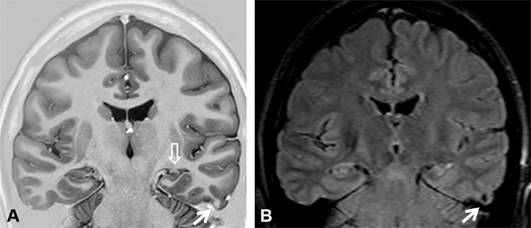
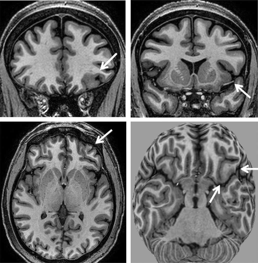
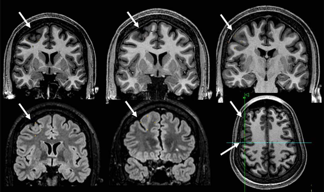
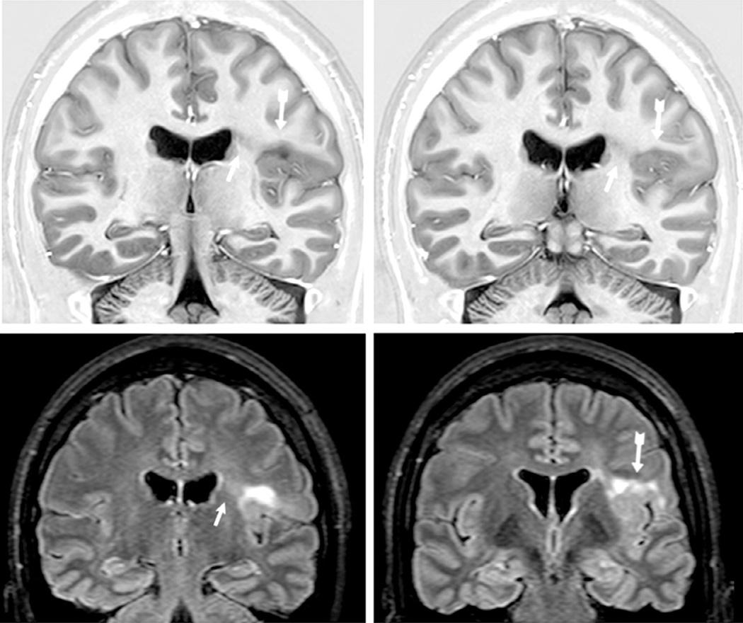
Imaging is pivotal in the evaluation and management of patients with seizure disorders. Elegant structural neuroimaging with magnetic resonance imaging (MRI) may assist in determining the etiology of focal epilepsy and demonstrating the anatomical changes associated with seizure activity. The high diagnostic yield of MRI to identify the common pathological findings in individuals with focal seizures including mesial temporal sclerosis, vascular anomalies, low-grade glial neoplasms and malformations of cortical development has been demonstrated. Positron emission tomography (PET) is the most commonly performed interictal functional neuroimaging technique that may reveal a focal hypometabolic region concordant with seizure onset. Single photon emission computed tomography (SPECT) studies may assist performance of ictal neuroimaging in patients with pharmacoresistant focal epilepsy being considered for neurosurgical treatment. This chapter highlights neuroimaging developments and innovations, and provides a comprehensive overview of the imaging strategies used to improve the care and management of people with epilepsy.

Keywords: 18F-fluorodeoxyglucose–positron emission tomography (18F-FDG-PET); Epilepsy; computed tomography (CT); drug-resistant focal epilepsy; ictal single photon emission computed tomography (SPECT); magnetic resonance imaging (MRI); surgical treatment of epilepsy.


Figures

Fig. 51.1
Fig. 51.1
Coronal T1 inversion recovery (A) and fluid-attenuated inversion recovery (FLAIR) axial (B) and coronal (C) magnetic resonance imaging (MRI) showing left hippocampal atrophy associated with changes in morphology and internal structure and hyperintense FLAIR signal (arrows) – all classic signs of hippocampal sclerosis on MRI that were confirmed on postoperative histopathology. Patient with left mesial temporal-lobe epilepsy and seizure-free after left amygdalohippocampectomy.
Fig. 51.2
Fig. 51.2
Coronal T1 inversion recovery (IR) (A) and fluid-attenuated inversion recovery (FLAIR) (B) magnetic resonance imaging showing left hippocampal atrophy associated with change in morphology and internal structure (open arrow) and hyperintense FLAIR signal. There is also a small nodular lesion which is slightly hyperintense in the T1 IR and hypointense in FLAIR images (arrows) in the third temporal gyrus that corresponded to a calcified lesion on computed tomography scan. Postoperative histopathology confirmed hippocampal sclerosis and a degenerated calcified neurocysticercosis nodule. Patient with left mesial temporal-lobe epilepsy and seizure-free after left anterior temporal lobe resection.
Fig. 51.3
Fig. 51.3
T1-weighted magnetic resonance imaging (MRI) multiplanar (MPR) and curvilinear reconstruction (bottom right) in a patient with frontal-lobe seizures and previous MRIs considered as normal. Note an area with abnormal gyri and slightly thickened cortex and blurred cortical-subcortical transition in the left lateral-basal frontal lobe (arrows). These abnormalities and the focal cortical-subcortical blurring became obvious in the curvilinear reconstructions in layers going from 4 to 8 mm deep from the surface of the brain (bottom right). The T2-weighted and fluid-attenuated inversion recovery images did not show abnormal signal (not shown here). These changes are suggestive of focal cortical dysplasia (FCD) type I or IIA.
Fig. 51.4
Fig. 51.4
Magnetic resonance imaging (MRI) multiplanar reconstruction (MPR) in a patient with frontal-lobe seizures due to focal cortical dysplasia (FCD) who had previous MRIs considered as negative. Top row shows reconstructed volumetric coronal T1-weighted images, and bottom row shows reconstructed volumetric fluid-attenuated inversion recovery (FLAIR) images and an axial reconstructed T1-weighted image (all with 1-mm thickness). The area with FCD in the left frontal lobe presents with slightly thickened cortex associated with abnormal gyri and cortical dimple (arrows). The FLAIR images did not show a clearly abnormal signal, except for a slightly blurred cortical-subcortical transition. The postoperative histopathology showed classic signs of FCD type IIA.
Fig. 51.5
Fig. 51.5
Coronal T1 inversion recovery and axial fluid-attenuated inversion recovery (FLAIR) images showing typical changes of focal cortical dysplasia type IIB (arrows) confirmed by postoperative histopathology in a patient with refractory focal seizures with temporal-insular semiology. Note area of cortical thickening and loss of sharpness of the cortical-subcortical transition and cortical-subcortical signal changes (increased FLAIR signal and decreased T1 signal) below the area of cortical thickening that extends toward the ventricle (“transmantle” sign).
Fig. 51.6
Fig. 51.6
Coronal T1 inversion recovery and fluid-attenuated inversion recovery (FLAIR) images showing typical changes of the bottom of the sulcus dysplasia (arrows). This is a focal cortical dysplasia (FCD) type IIB localized in the bottom of a (usually) deep sulcus with a mildly thickened cortex and hyperintense FLAIR signal, as seen in this patient with left frontal epilepsy. Histopathology showed FCD type IIB and the patient became seizure-free after surgery.
Fig. 51.7
Fig. 51.7
Coronal T1-weighted inversion recovery and T2-weighted images (top) and sagittal T1-weighted and fluid-attenuated inversion recovery images showing a small left anterior temporal encephalocele (arrows) and a left hippocampus with an abnormal shape and loss of internal structure (open arrow), in a patient with left temporal-lobe epilepsy.
Fig. 51.8
Fig. 51.8
Open-lips (A, B) and closed-lips (C, D) schizencephaly; bilateral perisylvian polymicrogyria (E, F).
Fig. 51.9
Fig. 51.9
Unilateral periventricular nodular heterotopia (PNH) (A: arrow) with polymicrogyria in the adjacent cortex; two different patients with bilateral PNH (B, C); a patient with PNH in the right temporal horn of the ventricle (D: arrow) and a large subcortical heterotopia extending to the posterior quadrant of the brain (E, F); three patients with different thickness of subcortical laminar heterotopia (double cortex), from thin and discontinuous bands (G) to continuous bands (H, I); three patients with different degrees of lissencephaly–agyria–pachygyria complex, from pachygyria (J), posterior agyria and anterior pachygyria (K), to diffuse lissencephaly (L).
Fig. 51.10
Fig. 51.10
Coronal T1-weighted inversion recovery, axial fluid-attenuated inversion recovery (FLAIR), and T1-weighted sagittal images in a patient with right hemimegalencephaly, more intense in the frontal region. Note the abnormal signal in the white matter of the affected hemisphere (hypointense on T1-weighted and hyperintense on FLAIR images), areas of pachygyria and hemispheric enlargement. Periventricular nodular heterotopia is also present (coronal image).
Fig. 51.11
Fig. 51.11
Multiple cortical tubers (arrows) in a fluid-attenuated inversion recovery image in a patient with tuberous sclerosis and epilepsy.
Fig. 51.12
Fig. 51.12
Axial T2-weighted (A), susceptibility-weighted imaging (SWI) (B) and postgadolinium T1-weighted (C) images, and coronal fluid-attenuated inversion recovery (D) and T1-weighted (E, F) images in a patient with Sturge–Weber syndrome and epilepsy. Note the dural and leptomeningeal angiomatosis involving the right occipital and posterior temporal regions (arrows) which are best seen in the SWI (B). There is an associated right hippocampal sclerosis (open arrows), and an atrophy of the right temporal-occipital region.
Fig. 51.13
Fig. 51.13
Coronal magnetic resonance imaging (MRI) showing ganglioglioma in three patients with temporal-lobe epilepsy and seizures not responding to antiepileptic drugss who became seizure-free after surgical resection of the lesion. (A) T1 inversion recovery image showing a small ganglioglioma in the right collateral sulcus (arrow) which was previously missed on an MRI without thin coronal cuts. (B) T2-weighted image showing a ganglioglioma in the left amygdala. (C) Axial T1-weighted and (D) coronal T2-weighted images showing a ganglioglioma in the left uncal region with cystic and solid components.
Fig. 51.14
Fig. 51.14
Axial fluid-attenuated inversion recovery (FLAIR) (A) and T1-weighted (B) images and postgadolinium coronal T1-weighted image (with a slight peritumoral enhancement (C) in a patient with temporal-lobe epilepsy due to oligodendroglioma in the right temporal horn of the ventricle adjacent to the hippocampus. The area with hypointense signal on FLAIR (arrow) and T1 images corresponds to a small calcification seen on computed tomography scan.
Fig. 51.15
Fig. 51.15
(A–C) T1 postgadolinium, T2-weighted coronal and axial fluid-attenuated inversion recovery images showing a dysembryoplastic neuroepithelial tumor (DNT) associated with focal cortical dysplasia (confirmed in the postoperative histopathology) in the right temporal lobe in a 23-year-old patient with refractory partial seizures since the age of 9 years, who became seizure-free after lesionectomy. Note a small area of intratumoral contrast enhancement (A: arrow), and the classic heterogeneous aspect of the tumor in all magnetic resonance imaging (sequences, sometimes given the appearance of small microcysts inside the lesion. (D–F) Coronal T2-weighted and sagittal T1-weighted images showing a DNT in the left temporal lobe in a patient with seizures since childhood who became seizure-free after lesionectomy.
Fig. 51.16
Fig. 51.16
Axial T1-weighted and double inversion recovery (DIR), and three-dimensional fluid-attenuated inversion recovery (FLAIR) curvilinear reconstruction showing a left superior frontal gyrus lesion with hyperintense FLAIR signal and blurred cortical-subcortical interface in a 6-year-old girl with refractory frontal-lobe epilepsy. Postoperative histopathology showed an angiocentric glioma (a rare grade I glioma).
Fig. 51.17
Fig. 51.17
Axial fluid-attenuated inversion recovery (FLAIR), susceptibility-weighted imaging (SWI), and T1-weighted images in a patient with a cavernoma (arrows) and right frontal epilepsy (top) and coronal T1-weighted inversion recovery and FLAIR images in a patient with left temporal-lobe epilepsy due to a cavernoma (bottom). Note the classic hypointense signal surrounding the lesion on FLAIR and T1-weighted images.
Fig. 51.18
Fig. 51.18
Axial and coronal images from four patients with perinatal insults and epilepsy. (A) and (E) show hemiatrophy (right hemisphere); (B) and (F), a large left middle cerebral artery infarct; (C) and (G), a bilateral occipital gliosis and atrophy; and (D) and (H), porencephaly in the territory of the left middle cerebral artery. Note that all four patients have severe hippocampal atrophy, and in (H), hyperintense T2 signal ipsilateral to the main lesion.
Fig. 51.19
Fig. 51.19
Fluid-attenuated inversion recovery (FLAIR) and T1-weighted images showing an area of gliosis (best seen in the FLAIR images) and ulegyria (best seen in the sagittal image) in the left occipital region in a patient with refractory occipital epilepsy. Postoperative histopathology showed an area of focal cortical dysplasia and a gliotic scar tissue.
Fig. 51.20
Fig. 51.20
Coronal fluid-attenuated inversion recovery and T1-weighted images showing a hypothalamic hamartoma (arrows) in a patient with gelastic seizures.
Fig. 51.21
Fig. 51.21
Axial T2-weighted (A), coronal T1-weighted inversion recovery (B), fluid-attenuated inversion recovery (FLAIR), axial (C), and coronal (D) images from two patients with Rasmussen’s encephalitis (A, B and C, D respectively) involving the right hemisphere. The progressive atrophy usually involves initially the insular-opercular regions, as seen in these patients. Sometimes, multiple cortical and subcortical foci of hyperintense FLAIR signal may be present, even in the initial stages of the disease, as in the patient shown in (C) and (D).
Fig. 51.22
Fig. 51.22
(A) Right temporal focal hypometabolism seen on 18F-fluorodeoxyglucose positron emission tomography in a patient with normal magnetic resonance imaging (MRI) scan and right temporal seizure onset on ictal video-electroencephalogram monitoring. (B) MRI in the same patient.
Fig. 51.23
Fig. 51.23
(A) 18F-FCWAY positron emission tomography (PET) scan showing reduced left temporal 5HT-1A receptor binding in a patient with a left temporal focus on ictal video-electroencephalogram monitoring. (B) 18F-fluorodeoxyglucose PET shows less clear focal hypometabolism in the same region.
Fig. 51.24
Fig. 51.24
Subtraction ictal SPECT co-registered to a magnetic resonance imaging head (SISCOM) shows a region of focal hyperperfusion over the right frontal head region in a patient with nonlesional extratemporal epilepsy of right frontal-lobe origin.
Fig. 51.25
Fig. 51.25
STATISCOM (statistical ictal SPECT coregistered to magnetic resonance imaging) shows a left medial temporal-lobe region of hyperperfusion in a patient with left temporal-lobe epilepsy.

References

    1. Ahnlide J-A, Rosén I, Lindén-Mickelsson Tech P, et al. Does SISCOM contribute to favorable seizure outcome after epilepsy surgery? Epilepsia. 2007;48:579–588. - PubMed
    1. Andermann F. Rasmussen’s syndrome. Stoneham, MA: Butterworth-Heinemann; 1991. Chronic encephalitis and epilepsy.
    1. Andersen AR. 99mTc-D, L-hexamethylene-propyleneamine oxime (99mTc-HMPAO): basic kinetic studies of a tracer of cerebral blood flow. Cerebrovasc Brain Metab Rev. 1989;1:288–318. - PubMed
    1. Arruda F, Cendes F, Andermann F, et al. Mesial atrophy and outcome after amygdalohippocampectomy or temporal lobe removal. Ann Neurol. 1996;40:446–450. - PubMed
    1. Banati RB, Goerres GW, Myers R, et al. [11C](R)-PK11195 positron emission tomography imaging of activated microglia in vivo in Rasmussen’s encephalitis. Neurology. 1999;53:2199–2203. - PubMed