. 2016 Sep;38(3):767-75.
doi: 10.3892/ijmm.2016.2665. Epub 2016 Jul 5.

The neutrophil elastase inhibitor, sivelestat, attenuates sepsis-related kidney injury in rats

Affiliations

The neutrophil elastase inhibitor, sivelestat, attenuates sepsis-related kidney injury in rats

Guofu Li et al. Int J Mol Med. 2016 Sep.

Abstract

Sepsis-induced acute kidney injury (AKI) represents a major cause of mortality in intensive care units. Sivelestat, a selective inhibitor of neutrophil elastase (NE), can attenuate sepsis-related acute lung injury. However, whether sivelestat can preserve kidney function during sepsis remains unclear. In this study, we thus examined the effects of sivelestat on sepsis-related AKI. Cecal ligation and puncture (CLP) was performed to induce multiple bacterial infection in male Sprague-Dawley rats, and subsequently, 50 or 100 mg/kg sivelestat were administered by intraperitoneal injection immediately after the surgical procedure. In the untreated rats with sepsis, the mean arterial pressure (MAP) and glomerular filtration rate (GFR) were decreased, whereas serum blood urea nitrogen (BUN) and neutrophil gelatinase-associated lipocalin (NGAL) levels were increased. We found that sivelestat promoted the survival of the rats with sepsis, restored the impairment of MAP and GFR, and inhibited the increased BUN and NGAL levels; specifically, the higher dose was more effective. In addition, sivelestat suppressed the CLP-induced macrophage infiltration, the overproduction of pro-inflammatory mediators (tumor necrosis factor‑α, interleukin-1β, high-mobility group box 1 and inducible nitric oxide synthase) and serine/threonine kinase (Akt) pathway activation in the rats. Collectively, our data suggest that the inhibition of NE activity with the inhibitor, sivelestat, is beneficial in ameliorating sepsis-related kidney injury.


Figures
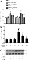
Figure 1
Figure 1
Sivelestat suppresses serum and kidney neutrophil elastase (NE) expression in septic rats. NE levels in (A) rat serum at 6 and 24 h and (B) kidney tissues at 24 h post-cecal ligation and puncture (CLP) surgery assessed using an ELISA kit (n=8 rats per group). (C) NE protein expression was detected by western blot analysis in rat kidney tissues at 24 h post-CLP procedure (n=5). β-actin was used as an endogenous control. Data are expressed as the means ± SD. No significant differences were observed in the sham-operated rats. ***P<0.001 compared with the sham-operated (Sham) group; P<0.05, †††P<0.001 compared with the sepsis group at the same time point.
Figure 2
Figure 2
Effects of sivelestat on kidney function in rats that underwent sham or cecal ligation and puncture (CLP) surgery. (A) Kaplan-Meier survival plots for all experimental rats (n=10 rats per group). ††P<0.01, compared with the sepsis group (log-rank test). (B) Rat mean arterial pressure (MAP) was detected at 24 h post-surgery (n=8–10 rats per group). (C) Representatives of hematoxylin and eosin (H&E) staining of rat kidney tissues. (D) Serum levels of blood urea nitrogen (BUN), (E) neutrophil gelatinase-associated lipocalin (NGAL) were determined at 6 and 24 h post-surgery using commercially available kits (n=8 rats per group). (F) Inulin clearance and (G) FENa were determined at 24 h post-surgery (n=8 rats per group). Data are expressed as the means ± SD. ***P<0.001 compared with the sham-operated (Sham) group; P<0.05, ††P<0.01, †††P<0.001 compared with the sepsis group at the same time point. Scale bars, 50 µm.
Figure 3
Figure 3
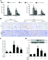
Sivelestat inhibits cecal ligation and puncture (CLP)-induced macrophage infiltration and pro-inflammation mediator release in rats. (A) Serum concentrations of tumor necrosis factor α (TNF-α) and interleukin-1β (IL-1β) were determined using the respective ELISA kits at 6 and 24 h after sham or CLP surgery (n=8 rats per group). (B) Macrophage infiltration in rat kidney tissues was assessed with specific antibody against ED-1 at 24 h after sham or CLP surgery (upper panels), and (C) the ED-1 positive cells were counted under a light microscope. (B) Representative images of immunohistochemical staining for high-mobility group box 1 (HMGB1) in rat kidney tissues at 24 h post-surgery (lower panels). (D) Renal HMGB1 protein expression was detected by western blot analysis (n=5). β-actin was used as an endogenous control. Data are expressed as the means ± SD. ***P<0.001 compared with the sham-operated (Sham) group; P<0.05, †††P<0.001 compared with the sepsis group at the same time point. Scale bars, 20 µm.
Figure 4
Figure 4
Sivelestat inhibits cecal ligation and puncture (CLP)-induced nitric oxide synthase (iNOS) upregulation in rat kidney tissues. (A) Representative images of immunohistochemical staining fir iNOS in rat kidney tissues at 24 h post-CLP procedure. (B) Renal iNOS protein expression was detected by western blot analysis (n=5). β-actin was used as an endogenous control. Data are expressed as the means ± SD. ***P<0.001 compared with the sham-operated (Sham) group; P<0.05, †††P<0.001 compared with the sepsis group. Scale bars, 20 µm.
Figure 5
Figure 5
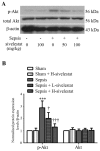
Sivelestat inhibits the cecal ligation and puncture (CLP)-induced activation of the phosphatidylinositol 3-kinase (PI3K)/Akt pathway activation in rat kidney tissues. (A) Representative blots for total Akt and phospho-Akt (p-Akt) in rat kidney tissues at 24 h post-CLP procedure (n=5). β-actin was used as an endogenous control. (B) Protein expression levels of total Akt and p-Akt were normalized as ratios to β-actin. Data are expressed as the means ± SD. ***P<0.001 compared with the sham-operated (Sham) group; P<0.05, †††P<0.001 compared with the sepsis group.

Similar articles

Cited by

References

    1. Fry DE. Sepsis, systemic inflammatory response, and multiple organ dysfunction: the mystery continues. Am Surg. 2012;78:1–8. - PubMed
    1. Rittirsch D, Flierl MA, Ward PA. Harmful molecular mechanisms in sepsis. Nat Rev Immunol. 2008;8:776–787. doi: 10.1038/nri2402. - DOI - PMC - PubMed
    1. Morrell ED, Kellum JA, Pastor-Soler NM, Hallows KR. Septic acute kidney injury: molecular mechanisms and the importance of stratification and targeting therapy. Crit Care. 2014;18:501. doi: 10.1186/s13054-014-0501-5. - DOI - PMC - PubMed
    1. Chen L, Yang S, Zumbrun EE, Guan H, Nagarkatti PS, Nagarkatti M. Resveratrol attenuates lipopolysaccharide-induced acute kidney injury by suppressing inflammation driven by macrophages. Mol Nutr Food Res. 2015;59:853–864. doi: 10.1002/mnfr.201400819. - DOI - PMC - PubMed
    1. Shimazu R, Akashi S, Ogata H, Nagai Y, Fukudome K, Miyake K, Kimoto M. MD-2, a molecule that confers lipopolysaccharide responsiveness on toll-like receptor 4. J Exp Med. 1999;189:1777–1782. doi: 10.1084/jem.189.11.1777. - DOI - PMC - PubMed

MeSH terms