

Differential Gene Expression Associated with Meningeal Injury in Acute Mild Traumatic Brain Injury
- PMID: 27430610
- PMCID: PMC5314966
- DOI: 10.1089/neu.2016.4479
Differential Gene Expression Associated with Meningeal Injury in Acute Mild Traumatic Brain Injury
Abstract
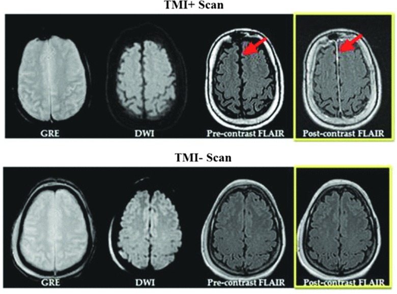
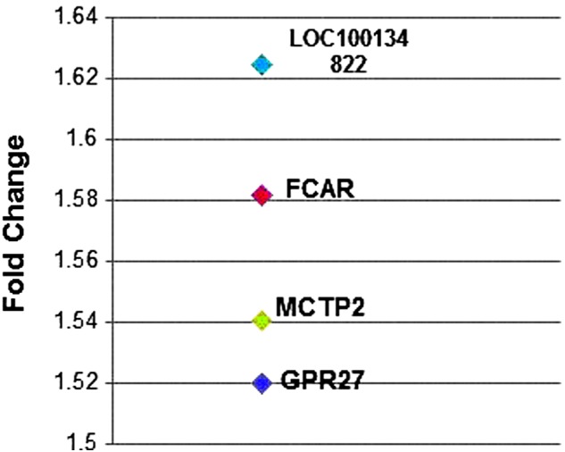
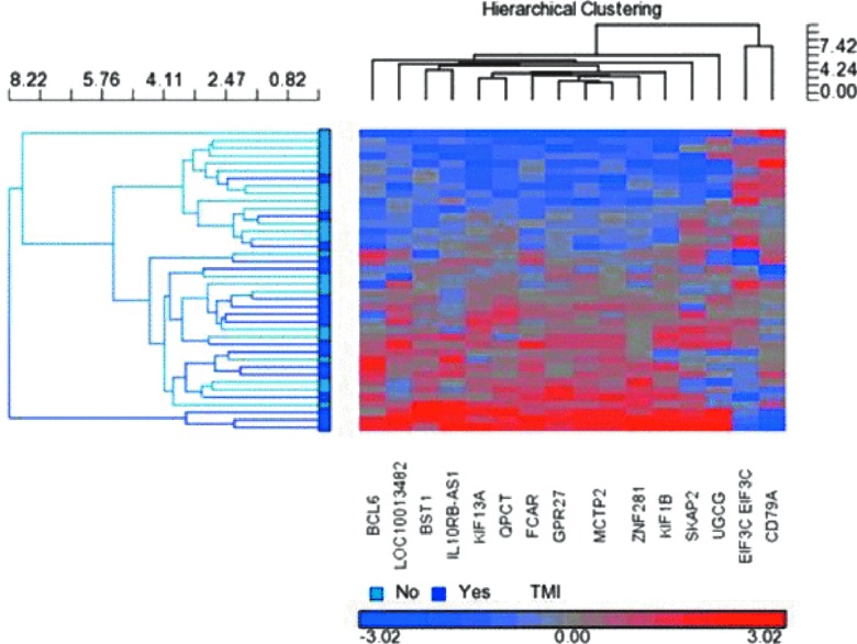
Injury to the meninges is not uncommon after traumatic brain injury (TBI), yet minimal research has been directed toward understanding the relevant biology. After a concussive event, the meninges are observed to abnormally enhance on post-contrast magnetic resonance imaging (MRI) in some patients, but not all. The aim of this work is to identify genes differentially expressed in patients with meningeal injury. Patients presenting to the emergency room with suspected TBI received a standard research MRI and blood draw within 48 h of injury. Two groups of patients were included: those with and without abnormal enhancement of the meninges on post-contrast MRI, both without other imaging findings. Groups were compared on microarray gene expression in peripheral blood samples using Affymetrix (Santa Clara, CA) and Partek Genomics Suite (Partek, Inc., St. Louis, MO) software (false discovery rate, <0.05). Forty patients were enrolled with a time from injury to MRI/blood draw of 16.8 h (interquartile range, 7.5-24.1). We observed 76 genes to be differentially expressed in patients with meningeal injury compared to those without, such as receptor for Fc fragment of IgA, multiple C2 domains, transmembrane 2, and G-protein-coupled receptor 27, which have been previously associated with initiating inflammatory mediators, phagocytosis, and other regulatory mechanisms. Post-contrast MRI is able to detect meningeal injury and has a unique biological signature observed through gene expression. These findings suggest that an acute inflammatory response occurs in response to injury to the meninges following a concussion.
Keywords: MRI; brain trauma; concussion; gene expression.
Conflict of interest statement
No competing financial interests exist.
Figures
References
-
- Michael D.B., Byers D.M., and Irwin L.N. (2005). Gene expression following traumatic brain injury in humans: analysis by microarray. J. Clin. Neurosci. 12, 284–290 - PubMed
-
- Grond-Ginsbach C., Hummel M., Wiest T., Horstmann S., Pfleger K., Hergenhahn M., Hollstein M., Mansmann U., Grau A.J., and Wagner S. (2008). Gene expression in human peripheral blood mononuclear cells upon acute ischemic stroke. J. Neurol. 255, 723–731 - PubMed
Publication types
MeSH terms
Grants and funding
LinkOut - more resources
Full Text Sources
Other Literature Sources
Medical
Miscellaneous
