. 2016 Aug 15;197(4):1287-97.
doi: 10.4049/jimmunol.1600266. Epub 2016 Jul 18.

HIF-1α Is an Essential Mediator of IFN-γ-Dependent Immunity to Mycobacterium tuberculosis

Affiliations

HIF-1α Is an Essential Mediator of IFN-γ-Dependent Immunity to Mycobacterium tuberculosis

Jonathan Braverman et al. J Immunol. .

Abstract

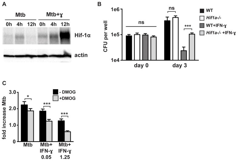
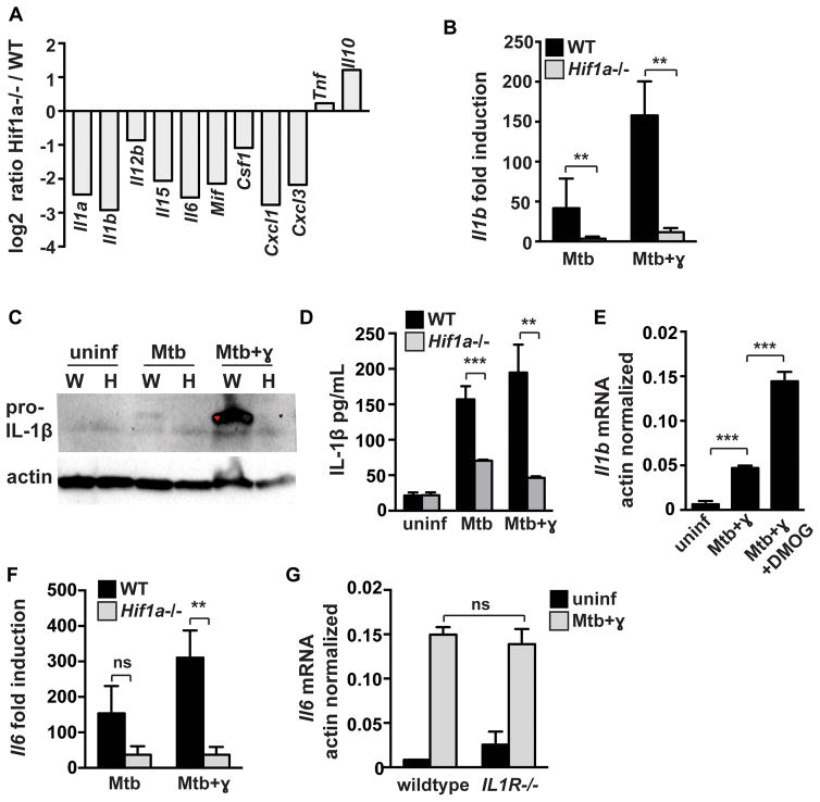
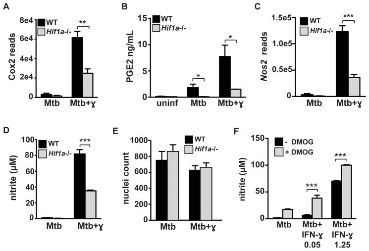
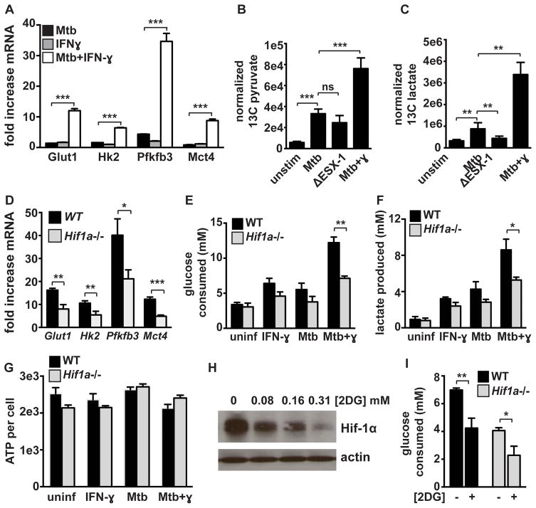
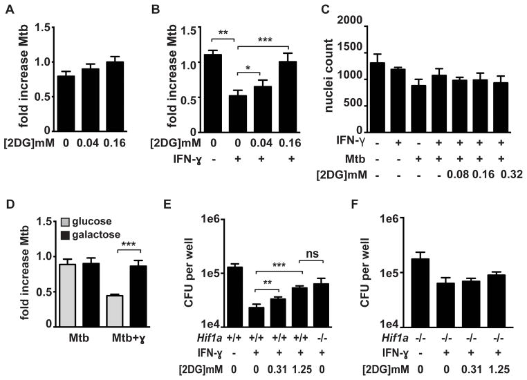
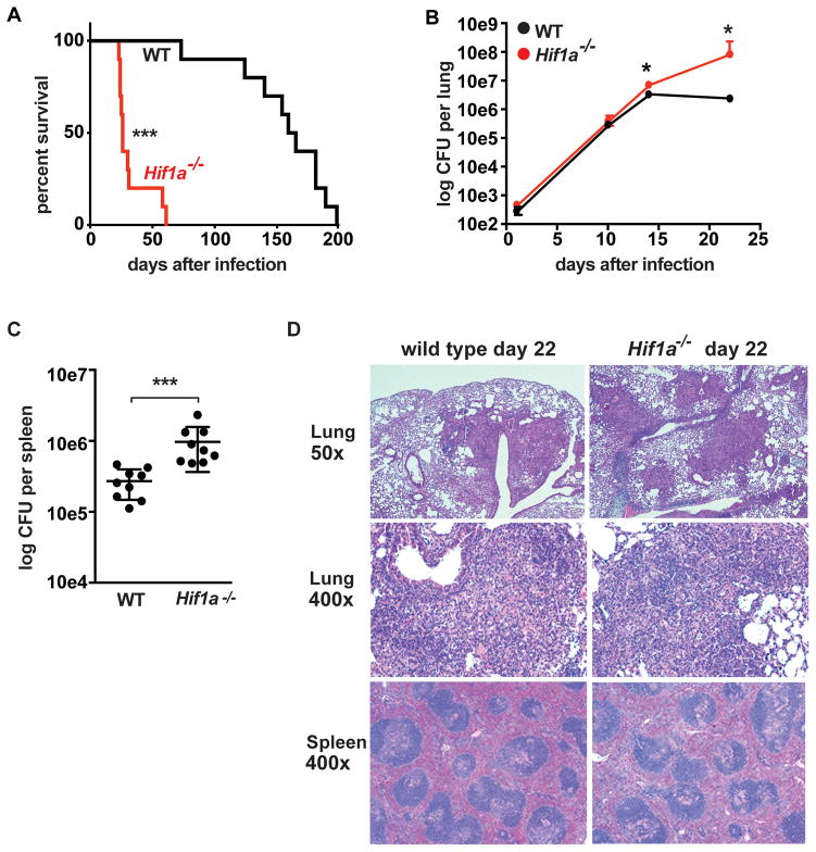
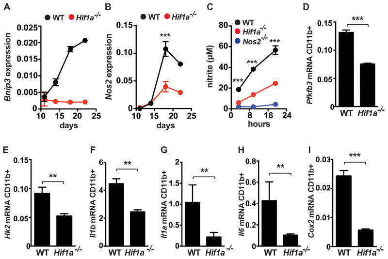
The cytokine IFN-γ coordinates macrophage activation and is essential for control of pathogens, including Mycobacterium tuberculosis However, the mechanisms by which IFN-γ controls M. tuberculosis infection are only partially understood. In this study, we show that the transcription factor hypoxia-inducible factor-1α (HIF-1α) is an essential mediator of IFN-γ-dependent control of M. tuberculosis infection both in vitro and in vivo. M. tuberculosis infection of IFN-γ-activated macrophages results in a synergistic increase in HIF-1α protein levels. This increase in HIF-1α levels is functionally important, as macrophages lacking HIF-1α are defective for IFN-γ-dependent control of infection. RNA-sequencing demonstrates that HIF-1α regulates nearly one-half of all IFN-γ-inducible genes during infection of macrophages. In particular, HIF-1α regulates production of important immune effectors, including inflammatory cytokines and chemokines, eicosanoids, and NO. In addition, we find that during infection HIF-1α coordinates a metabolic shift to aerobic glycolysis in IFN-γ-activated macrophages. We find that this enhanced glycolytic flux is crucial for IFN-γ-dependent control of infection in macrophages. Furthermore, we identify a positive feedback loop between HIF-1α and aerobic glycolysis that amplifies macrophage activation. Finally, we demonstrate that HIF-1α is crucial for control of infection in vivo as mice lacking HIF-1α in the myeloid lineage are strikingly susceptible to infection and exhibit defective production of inflammatory cytokines and microbicidal effectors. In conclusion, we have identified HIF-1α as a novel regulator of IFN-γ-dependent immunity that coordinates an immunometabolic program essential for control of M. tuberculosis infection in vitro and in vivo.


Figures

Figure 1
Figure 1. HIF-1α is required for IFN-γ based control of M. tuberculosis replication in macrophages
(A) Timecourse of HIF-1α protein stabilization by western blotting after infection of resting and IFN-γ activated WT BMDM with M. tuberculosis at MOI=5 with the 0h timepoint reflecting the end of the 4h phagocytosis period. (B) WT and Hif1a−/− BMDM were infected with M. tuberculosis at MOI=5 and bacterial replication was monitored with and without IFN-γ treatment by plating for CFU. CFU on day 0 and day 3 are shown. (C) Resting and IFN-γ activated WT BMDM were infected with the TB-lux strain of M. tuberculosis and treated with 200 μM DMOG after phagocytosis of bacteria. Bacterial growth was assessed by reading RLU immediately after phagocytosis and at 72h after infection and fold change is shown. For all experiments error bars represent the standard deviation of a minimum of quadruplicate wells and a representative experiment of a minimum of 3 is shown. p-values were determined using an unpaired t-test. ***p≤0.001, *p≤0.05.
Figure 2
Figure 2. HIF-1α regulates cytokine and chemokine production
Resting and IFN-γ activated WT and Hif1a−/− BMDM were infected with M. tuberculosis at MOI=5. RNA was prepared for RNA-seq at 24h post infection. (A) RNA-seq data showing transcript levels of cytokines and chemokines in Hif1a−/− BMDM relative to WT BMDM on a log2 scale. Data shown is from macrophages treated with IFN-γ and infected with M. tuberculosis. (B) RNA-seq data showing fold induction of Il1b transcript over untreated macrophages in WT and Hif1a−/− BMDM (C) Western blotting for pro-IL-1b from cell lysates at 12h after infection in wild-type [W] and Hif1a−/− [H] macrophages. (D) ELISA for IL1b from cell supernatants at 36h after infection. (E) qPCR data showing actin normalized Il1b transcript in WT BMDM with DMOG treatment. (F) RNAseq data showing fold induction of Il6 transcript over untreated macrophages in WT and Hif1a−/− BMDM (G) qPCR data showing actin normalized Il6 transcript levels in WT and IL1R deficient BMDM. For RNA-seq, RNA was prepared from 3 independent infections. All other experiments were repeated 2–3 times and representative experiments are shown. Error bars represent standard deviation. p values were determined using an unpaired t-test. ***p≤0.001, **p≤0.01, *p≤0.05.
Figure 3
Figure 3. HIF-1α is required for full activation of IFN-γ dependent cell intrinsic immune responses
Resting and IFN-γ activated WT and Hif1a−/− BMDM were infected with M. tuberculosis at MOI=5. RNA was prepared for RNA-seq at 24h post infection. (A) RNA-seq data showing expression levels of Cox2 (official name Ptgs2) in WT and Hif1a−/− BMDM. (B) PGE2 ELISA from macrophage supernatants at 36h after infection. (C) RNA-seq data showing expression levels of Nos2 in WT and Hif1a−/− BMDM (D) Griess assay for nitric oxide production in WT and Hif1a−/− BMDM. (E) Nuclei counts after DAPI staining in WT and Hif1a−/− BMDM 24h after infection. (F) Resting and IFN-γ activated BMDM were infected with M. tuberculosis and treated with 200 μM DMOG after phagocytosis of bacteria and a Griess assay was performed 72h after infection. For RNA-seq, RNA was prepared from 3 independent infections. All other experiments are representative of 3 or more. Error bars represent standard deviation. p-values were determined using an unpaired t-test. ***p≤0.001, **p≤0.01, *p≤0.05.
Figure 4
Figure 4. HIF-1α mediates the transition to aerobic glycolysis in M. tuberculosis infected macrophages but is not required for maintenance of ATP production
(A) Fold increase in transcript levels over resting BMDM for the glycolytic genes Glut1, Hk2, Pfkfb3, and Mct4 were measured by qPCR in WT BMDM. (B and C) BMDM infected with M. tuberculosis were cultured in media containing 13C labeled glucose for 24h at which time lysates were prepared and 13C labeled pyruvate (B) and lactate (C) were detected by LC-MS/MS. Values represent the average of quintuplicate wells and error is standard deviation. (D) RNAseq data showing fold increase over resting BMDM of glycolytic genes in WT and Hif1a−/− BMDM treated with IFN-γ and infected with M. tuberculosis. (E) Glucose consumption after 36h by WT and Hif1a−/− BMDM. (F) Lactate production after 24h by WT and Hif1a−/− BMDM. (G) ATP levels were measured using a luciferase based assay (CellTiter-Glo) and were normalized to cell number measured by DAPI staining and counting of nuclei. (H) HIF-1α western blot 12h after M. tuberculosis infection of IFN-γ treated BMDM, treated with increasing dose of 2-DG following 4h phagocytosis period. (I) Glucose consumption 24h post infection of WT and Hif1a−/− BMDM with and without 2DG treatment. Data in (A),(D),(E),(F) and (H) are representative of 3 or more experiments, data in (G) and (I) are representative of 2 experiments. Error bars represent standard deviation. p-values were determined using an unpaired t-test. ***p≤0.001, **p≤0.01, *p≤0.05
Figure 5
Figure 5. Enhanced flux through glycolysis is required for IFN-γ dependent control of M. tuberculosis infection
Resting (A) and IFN-γ activated (B) BMDM were infected with the TB-lux strain of M. tuberculosis and treated with 2DG immediately after the 4h phagocytosis period. Bacterial growth was assessed by reading RLU immediately after phagocytosis and at 24h after infection and fold change is shown. (C) Macrophage viability was assessed using DAPI staining of nuclei and microscopy 24h after infection. (D) Resting and IFN-γ activated macrophages were infected with the TB-lux strain of M. tuberculosis and were switched to glucose free media containing galactose immediately after the 4h phagocytosis period and fold growth was determined at 24h after infection. (E and F) WT and Hif1a−/− BMDM were infected with M. tuberculosis at MOI=5 and bacterial replication was monitored by plating for CFU. 2DG treatment at the indicated concentrations began after the 4h phagocytosis and 2DG was washed out 24h later. CFU 72h post infection is shown. Representative experiments of 3 or more are shown for (A–D) and representative of 2 experiments for (E) and (F). Error bars are standard deviation from 3–6 replicate wells for TB-lux data, 3 replicate wells for nuclei counts, and 5 replicate wells for CFU. p-values were determined using an unpaired t-test, ***p≤0.001, **p≤0.01, *p≤0.05
Figure 6
Figure 6. HIF-1α is required for control of M. tuberculosis infection in vivo
WT and Hif1a−/− mice on the C57BL/6 background were infected with ~400 CFU of the virulent M. tuberculosis strain Erdman via the aerosol route. (A) Survival following aerosol infection is shown for WT and Hif1a−/− mice. Experiment shown is representative of three experiments using 10–12 mice per genotype. (B) Bacterial loads in the lungs of WT and Hif1a−/− mice were enumerated by plating for CFU. Timecourse is representative of 3 experiments with 4–5 mice used per group at each timepoint. (C) Bacterial loads in the spleens at 21d after infection. Data from two pooled experiments are shown. (D) Lung tissues were fixed, embedded in paraffin, sectioned, and stained with haematoxylin and eosin. Images were obtained at 50x and 400x magnification. Error bars represent standard deviation. For survival curves p values were determined using the Mantel-Cox log. For CFU, p-values were determined using Mann-Whitney U. ***p<0.001, *p≤0.05.
Figure 7
Figure 7. HIF-1α regulates immune effectors in vivo
WT and Hif1a−/− mice on the C57BL/6 background were infected with ~400 CFU of the virulent M. tuberculosis strain Erdman via the aerosol route. CD11b+ cells were isolated from lungs of infected mice at the indicated timepoints and analyzed by qPCR for expression of Bnip3 (A) and Nos2 (B). (C) isolated CD11b+ cells were plated and the supernatants were assayed for nitric oxide production by Griess assay at the indicated timepoints. (D–I) qPCR from isolated from CD11b+ cells at 21 days post-infection in WT and Hif1a−/− mice. Data shown in (C–I) is represtentative of 2–3 experiments. p-values were determined using an unpaired t-test, ***p≤0.001, **p≤0.01.

References

    1. Floyd K. World Health Organization, editor. Global Tuberculosis Report 2012. WHO Press; 2012. pp. 1–100.
    1. Abel L, Casanova JL. Genetic predisposition to clinical tuberculosis: bridging the gap between simple and complex inheritance. American journal of human genetics. 2000;67:274. - PMC - PubMed
    1. Flynn JL, Chan J, Triebold KJ, Dalton DK, Stewart TA, Bloom BR. An essential role for interferon gamma in resistance to Mycobacterium tuberculosis infection. J Exp Med. 1993;178:2249–2254. - PMC - PubMed
    1. Cooper AM, Dalton DK, Stewart TA, Griffin JP, Russell DG, Orme IM. Disseminated tuberculosis in interferon gamma gene-disrupted mice. J Exp Med. 1993;178:2243–2247. - PMC - PubMed
    1. Zhang YJ, Reddy MC, Ioerger TR, Rothchild AC, Dartois V, Schuster BM, Trauner A, Wallis D, Galaviz S, Huttenhower C, Sacchettini JC, Behar SM, Rubin EJ. Tryptophan biosynthesis protects mycobacteria from CD4 T-cell-mediated killing. Cell. 2013;155:1296–1308. - PMC - PubMed

Publication types

Substances