

Review
doi: 10.1007/s11427-016-5088-4.
Epub 2016 Jul 19.
Binding of transition metals to S100 proteins
Affiliations
- PMID: 27430886
- PMCID: PMC5123432
- DOI: 10.1007/s11427-016-5088-4
Item in Clipboard
Review
Binding of transition metals to S100 proteins
Sci China Life Sci.
2016 Aug.
Abstract
The S100 proteins are a unique class of EF-hand Ca(2+) binding proteins distributed in a cell-specific, tissue-specific, and cell cycle-specific manner in humans and other vertebrates. These proteins are distinguished by their distinctive homodimeric structure, both intracellular and extracellular functions, and the ability to bind transition metals at the dimer interface. Here we summarize current knowledge of S100 protein binding of Zn(2+), Cu(2+) and Mn(2+) ions, focusing on binding affinities, conformational changes that arise from metal binding, and the roles of transition metal binding in S100 protein function.
Keywords: Copper; Manganese; S100 Proteins; Zinc.
Figures
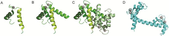
Structural features of S100 proteins. Ribbon diagrams of (A) an EF-hand motif, (B) an EF-hand domain, (C) the integration of two EF-hand domains into an S100 dimer, and (D) the alternate arrangement of two EF-hand domains in a prototypical EF-hand Ca2+ signal modulator. S100A12 was selected as the representative member of the S100 proteins and panels A, B and C were created using the Ca2+-loaded protein (PDB entry 1E8A). Panel D was created using Ca2+-loaded calmodulin (PDB entry 1CLL). The compact nature of the S100 homodimer relative to calmodulin implies a fundamentally different structural mechanism for transduction of Ca2+ signals.
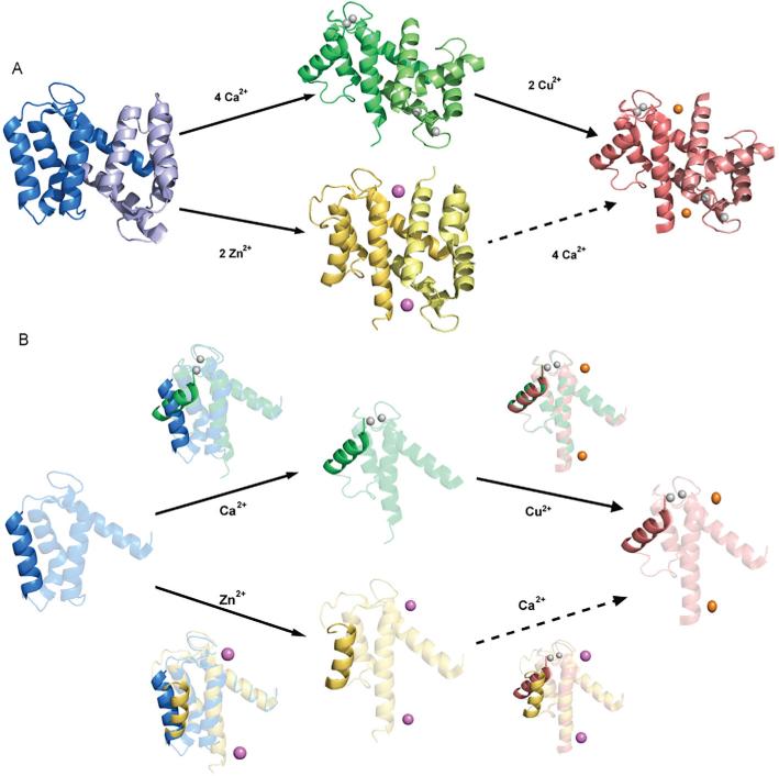
Three-dimensional structure of S100A12 and conformational changes induced by Ca2+ and transition metals. A, Ribbon diagrams of the apo, (Ca2+)4, (Zn2+)2, and (Ca2+)4, (Cu2+)2, states. B, Comparison of single sub-units to emphasize the differences in the packing of Helix III in different states. This reveals that the consequences of binding Ca2+ are much greater than those of binding transition metals. Images generated in pymol (DeLano, 2002) using coordinates deposited in the PDB for apo (2WCF), (Ca2+)4 (1E8A), (Zn2+)2 (2WC8) and (Ca2+)4, (Cu2+)2 (1ODB).

Alignments of S100 proteins containing transition metal binding sites. Structure based sequence alignment of S100 proteins from the His-rich (upper panel) and Cys-rich (lower panel) categories. Conserved residues in His-rich sites are highlighted with teal background, and those in Cys-rich sites in red background. Note the high degree of conservation in the His-rich proteins compared to the Cys-rich proteins. The alignments were generated using PROMALS3D (Pei et al., 2008).
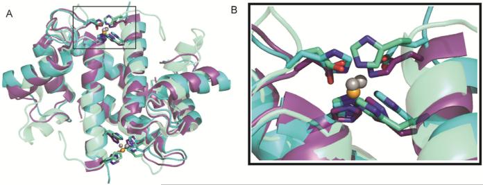
Structural similarity of tetrahedral zinc and copper binding sites in S100 proteins. A, Overlay of the structures of (Ca2+)4, (Zn2+)2-S100A7 (light green), (Ca2+)4, (Zn2+)2-S100B (teal) and (Ca2+)4,(Cu2+)2-S100A12 (purple) showing that the transition metal ions are chelated in a similar manner by side chains in the same position in the sequence. B, Zoom in on the tetrahedral Zn2+ and Cu2+ sites showing the similar spatial disposition of the 3 His and 1 Asp chelating side chains. The Zn2+ and Cu2+ ions are colored gray and orange, respectively. Images generated in pymol (DeLano, 2002) using PDB coordinates deposited for (Ca2+)4, (Zn2+)2-S100A7 (2PSR), (Ca2+)4, (Zn2+)2-S100B (3D0Y), and (Ca2+)4, (Cu2+)2-S100A12 (1ODB).
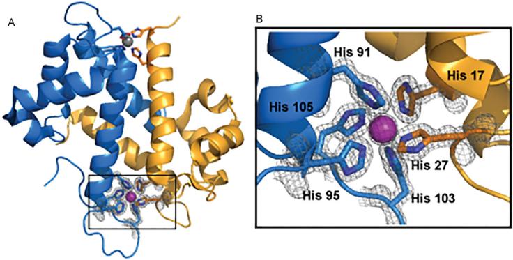
Three-dimensional structure of calprotectin highlighting the unique manganese binding site. The S100A8 subunit is colored blue, the S100A9 subunit gold, and the Mn2+ ion purple. An additional low occupancy Mn2+ ion is shown in gray. Images generated in pymol (DeLano, 2002) using coordinates deposited in the PDB for (Ca2+)4, (Mn2+)-CP (4GGF).
References
-
- Arnesano F, Banci L, Bertini I, Fantoni A, Tenori L, Viezzoli MS. Structural interplay between calcium(II) and copper(II) binding to S100A13 protein. Angew Chem Int Ed Engl. 2005;44:6341–6344. - PubMed
-
- Baudier J, Glasser N, Gerard D. Ions binding to s100 proteins. I. Calcium- and zinc-binding properties of bovine brain s100 alpha alpha, s100a (alpha beta), and s100b (beta beta) protein: Zn2+ regulates Ca2+ binding on s100b protein. J Biol Chem. 1986;261:8192–8203. - PubMed
-
- Baudier J, Glasser N, Haglid K, Gerard D. Purification, characterization and ion binding properties of human brain S100b protein. Biochim Biophys Acta. 1984;790:164–173. - PubMed
-
- Baudier J, Holtzscherer C, Gerard D. Zinc-dependent affinity chromatography of the S100b protein on phenyl-Sepharose. A rapid purification method. FEBS Lett. 1982;148:231–234. - PubMed
-
- Bhattacharya S, Bunick CG, Chazin WJ. Target selectivity in EF-hand calcium binding proteins. Biochim Biophys Acta. 2004;1742:69–79. - PubMed
Publication types
MeSH terms
Substances
Grants and funding
LinkOut - more resources
Full Text Sources
Other Literature Sources
Miscellaneous
