

Labeling mesenchymal cells with DMSA-coated gold and iron oxide nanoparticles: assessment of biocompatibility and potential applications
- PMID: 27431051
- PMCID: PMC4949766
- DOI: 10.1186/s12951-016-0213-x
Labeling mesenchymal cells with DMSA-coated gold and iron oxide nanoparticles: assessment of biocompatibility and potential applications
Abstract
Background: Nanoparticles' unique features have been highly explored in cellular therapies. However, nanoparticles can be cytotoxic. The cytotoxicity can be overcome by coating the nanoparticles with an appropriated surface modification. Nanoparticle coating influences biocompatibility between nanoparticles and cells and may affect some cell properties. Here, we evaluated the biocompatibility of gold and maghemite nanoparticles functionalized with 2,3-dimercaptosuccinic acid (DMSA), Au-DMSA and γ-Fe2O3-DMSA respectively, with human mesenchymal stem cells. Also, we tested these nanoparticles as tracers for mesenchymal stem cells in vivo tracking by computed tomography and as agents for mesenchymal stem cells magnetic targeting.
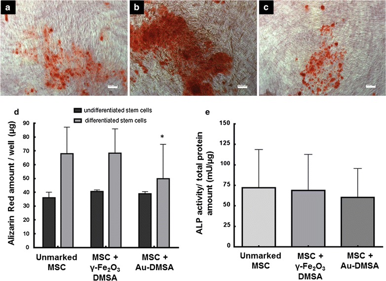
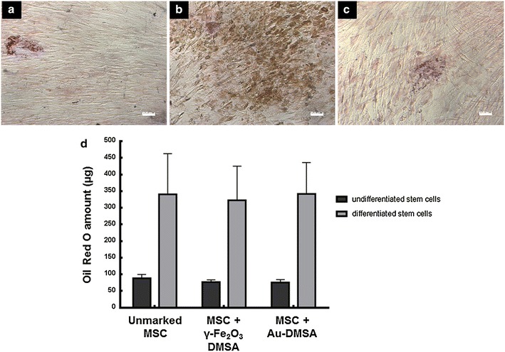
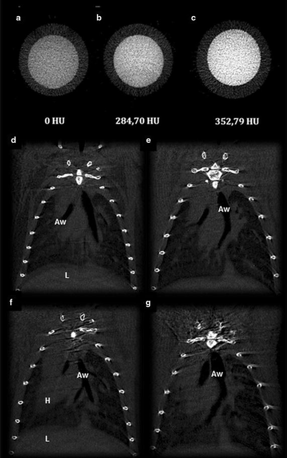
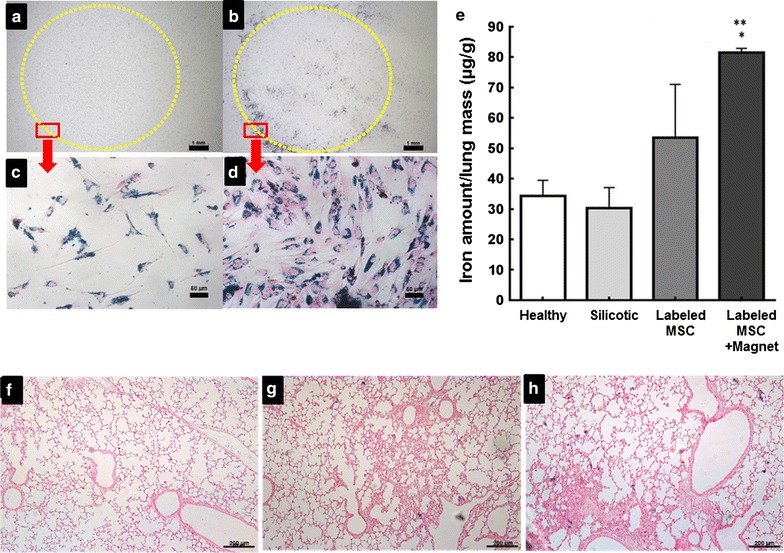
Results: Significant cell death was not observed in MTT, Trypan Blue and light microscopy analyses. However, ultra-structural alterations as swollen and degenerated mitochondria, high amounts of myelin figures and structures similar to apoptotic bodies were detected in some mesenchymal stem cells. Au-DMSA and γ-Fe2O3-DMSA labeling did not affect mesenchymal stem cells adipogenesis and osteogenesis differentiation, proliferation rates or lymphocyte suppression capability. The uptake measurements indicated that both inorganic nanoparticles were well uptaken by mesenchymal stem cells. However, Au-DMSA could not be detected in microtomograph after being incorporated by mesenchymal stem cells. γ-Fe2O3-DMSA labeled cells were magnetically responsive in vitro and after infused in vivo in an experimental model of lung silicosis.
Conclusion: In terms of biocompatibility, the use of γ-Fe2O3-DMSA and Au-DMSA as tracers for mesenchymal stem cells was assured. However, Au-DMSA shown to be not suitable for visualization and tracking of these cells in vivo by standard computed microtomography. Otherwise, γ-Fe2O3-DMSA shows to be a promising agent for mesenchymal stem cells magnetic targeting.
Keywords: Biocompatibility; Computed microtomography; DMSA-nanoparticles; Gold nanoparticles; Iron oxide nanoparticle; Magnetic targeting; Mesenchymal stem cells.
Figures


Similar articles
-
Detection of human breast cancer cells using a 2-deoxy-D-glucose-functionalized superparamagnetic iron oxide nanoparticles.Cancer Biomark. 2017;18(4):367-374. doi: 10.3233/CBM-160258. Cancer Biomark. 2017. PMID: 28106540
-
MRI of iron oxide nanoparticle-labeled ADSCs in a model of hindlimb ischemia.Biomaterials. 2013 Jul;34(21):4914-25. doi: 10.1016/j.biomaterials.2013.03.014. Epub 2013 Mar 25. Biomaterials. 2013. PMID: 23535037
-
[Comparison of the targeting properties of 2-deoxy-D-glucose-conjugated nanoparticles to breast cancer MDA-MB-231 cells and breast fibroblasts cells].Zhonghua Zhong Liu Za Zhi. 2013 Aug;35(8):566-71. Zhonghua Zhong Liu Za Zhi. 2013. PMID: 24314212 Chinese.
-
Poly(N,N-dimethylacrylamide)-coated maghemite nanoparticles for labeling and tracking mesenchymal stem cells.2009 Dec 23 [updated 2010 Feb 16]. In: Molecular Imaging and Contrast Agent Database (MICAD) [Internet]. Bethesda (MD): National Center for Biotechnology Information (US); 2004–2013. 2009 Dec 23 [updated 2010 Feb 16]. In: Molecular Imaging and Contrast Agent Database (MICAD) [Internet]. Bethesda (MD): National Center for Biotechnology Information (US); 2004–2013. PMID: 20641410 Free Books & Documents. Review.
-
The application of super paramagnetic iron oxide-labeled mesenchymal stem cells in cell-based therapy.Mol Biol Rep. 2013 Mar;40(3):2733-40. doi: 10.1007/s11033-012-2364-7. Epub 2012 Dec 27. Mol Biol Rep. 2013. PMID: 23269616 Review.
Cited by
-
Modulation of fibronectin and laminin expression by Rhodium (II) citrate-coated maghemite nanoparticles in mice bearing breast tumor.Sci Rep. 2017 Dec 20;7(1):17904. doi: 10.1038/s41598-017-18204-1. Sci Rep. 2017. PMID: 29263369 Free PMC article.
-
Recent trends in preparation and biomedical applications of iron oxide nanoparticles.J Nanobiotechnology. 2024 Jan 8;22(1):24. doi: 10.1186/s12951-023-02235-0. J Nanobiotechnology. 2024. PMID: 38191388 Free PMC article. Review.
-
Nanotechnology bring a new hope for asthmatics.Ann Transl Med. 2019 Oct;7(20):516. doi: 10.21037/atm.2019.09.153. Ann Transl Med. 2019. PMID: 31807498 Free PMC article. No abstract available.
-
Evaluation of Dimercaptosuccinic Acid-Coated Iron Nanoparticles Immunotargeted to Amyloid Beta as MRI Contrast Agents for the Diagnosis of Alzheimer's Disease.Cells. 2023 Sep 14;12(18):2279. doi: 10.3390/cells12182279. Cells. 2023. PMID: 37759500 Free PMC article.
-
Vascular Repair by Grafting Based on Magnetic Nanoparticles.Pharmaceutics. 2022 Jul 8;14(7):1433. doi: 10.3390/pharmaceutics14071433. Pharmaceutics. 2022. PMID: 35890328 Free PMC article. Review.
References
-
- Nikalje AP. Nanotechnology and its Applications in Medicine. Med chem. 2015;5:081–089. doi: 10.4172/2161-0444.1000247. - DOI
MeSH terms
Substances
LinkOut - more resources
Full Text Sources
Other Literature Sources
