

Evaluation of oral keratinocyte progenitor and T-lymphocite cells response during early healing after augmentation of keratinized gingiva with a 3D collagen matrix - a pilot study
- PMID: 27431208
- PMCID: PMC4948093
- DOI: 10.1186/s12903-016-0240-x
Evaluation of oral keratinocyte progenitor and T-lymphocite cells response during early healing after augmentation of keratinized gingiva with a 3D collagen matrix - a pilot study
Abstract
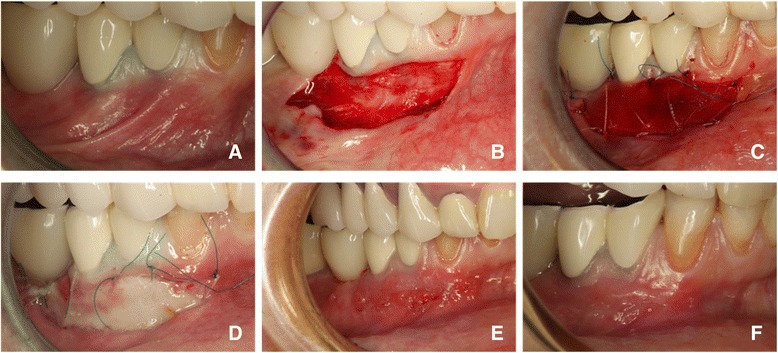
Background: The aim of the present study is to analyze the behavior of selected populations of oral keratinocytes and T-lymphocytes, responsible for re-constructing and maintaining the oral epithelial tissue architecture, following augmentation of the keratinized oral mucosa using a 3D-collagen matrix.
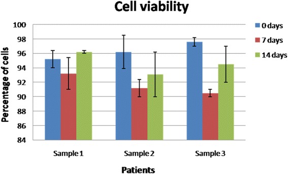
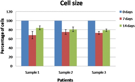
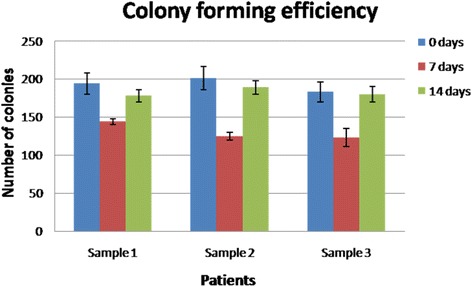
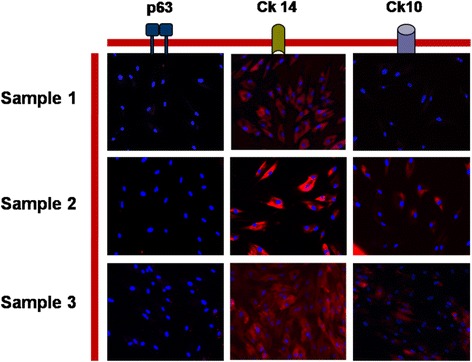
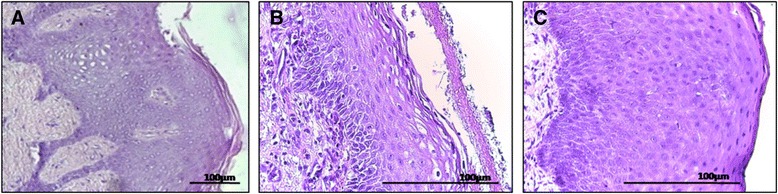
Methods: Different groups of oral keratinocytes were isolated from biopsies harvested from 3 patients before the surgical procedure, as well as 7 and 14 days after the augmentation procedure. T-lymphocytes were isolated from peripheral blood at same timepoints. Keratinocytes were characterized for stem and differentiation markers, such as p63, cytokeratin 10 and 14, and in vitro parameters, such as cell viability, cell size and colony-forming efficiency. T-lymphocytes were analyzed for viability and the expression of various cluster of differentiation markers. The methods included magnetic separation of cell populations, immunofluorescence, flow cytometry, and histology of oral biopsies.
Results: Both at 7 and 14 days, the majority of cells that repopulate the matrix were actively proliferating/progenitor oral keratinocytes with the phenotype integrin alfa6beta4 + CD71+. These cells display in vitro characteristics similar to the progenitor cells analyzed before the matrix placement. T-lymphocytes expressed CD8 and CD69 markers, while CD25 was absent.
Conclusion: The study shows that two weeks after the collagen membrane placement, the healing process appeared to be histologically complete, with no abnormal immune response induced by the matrix, however, with a higher than usual content of active proliferating cells, the majority of keratinocytes being characterized as transit amplifying cells.
Keywords: Collagen matrix; Keratinocytes; Oral mucosa; T-lymphocytes.
Figures
Similar articles
-
Novel method for proliferation of oral keratinocyte stem cells.J Periodontal Res. 2014 Dec;49(6):711-8. doi: 10.1111/jre.12153. Epub 2013 Dec 11. J Periodontal Res. 2014. PMID: 24329011
-
Interaction between a 3D collagen matrix used for periodontal soft tissue regeneration and T-lymphocytes: An in vitro pilot study.Exp Ther Med. 2019 Feb;17(2):990-996. doi: 10.3892/etm.2018.6979. Epub 2018 Nov 16. Exp Ther Med. 2019. PMID: 30679964 Free PMC article.
-
Development of a Full-Thickness Human Gingiva Equivalent Constructed from Immortalized Keratinocytes and Fibroblasts.Tissue Eng Part C Methods. 2016 Aug;22(8):781-91. doi: 10.1089/ten.TEC.2016.0066. Tissue Eng Part C Methods. 2016. PMID: 27406216 Free PMC article.
-
3D engineered human gingiva fabricated with electrospun collagen scaffolds provides a platform for in vitro analysis of gingival seal to abutment materials.PLoS One. 2022 Feb 3;17(2):e0263083. doi: 10.1371/journal.pone.0263083. eCollection 2022. PLoS One. 2022. PMID: 35113915 Free PMC article.
-
[Influencing factors for keratinized differentiation of keratinocytes].Zhonghua Kou Qiang Yi Xue Za Zhi. 2018 Apr 9;53(4):284-288. doi: 10.3760/cma.j.issn.1002-0098.2018.04.014. Zhonghua Kou Qiang Yi Xue Za Zhi. 2018. PMID: 29690702 Review. Chinese.
Cited by
-
Does Photobiomodulation Affects CK10 and CK14 in Oral Mucositis Radioinduced Repair?Int J Mol Sci. 2022 Dec 9;23(24):15611. doi: 10.3390/ijms232415611. Int J Mol Sci. 2022. PMID: 36555260 Free PMC article.
References
-
- Nevins M, Nevins ML, Kim S-W, Schupbach P, Kim DM. The use of mucograft collagen matrix to augment the zone of keratinized tissue around teeth: a pilot study. Int J Periodontics Restorative Dent. 2010;31(4):367–73. - PubMed
-
- Hall W, Lundergan W. Free gingival grafts. Current indications and techniques. Dent Clin N Am. 1993;37(2):227–42. - PubMed
MeSH terms
Substances
LinkOut - more resources
Full Text Sources
Other Literature Sources
Research Materials
