

Isolation of Mouse Hair Follicle Bulge Stem Cells and Their Functional Analysis in a Reconstitution Assay
- PMID: 27431247
- PMCID: PMC7450485
- DOI: 10.1007/978-1-4939-3786-8_8
Isolation of Mouse Hair Follicle Bulge Stem Cells and Their Functional Analysis in a Reconstitution Assay
Abstract
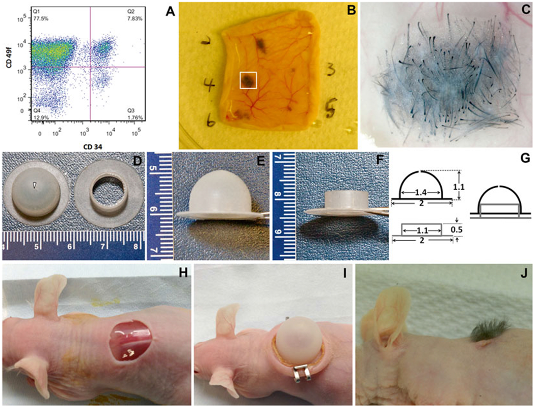
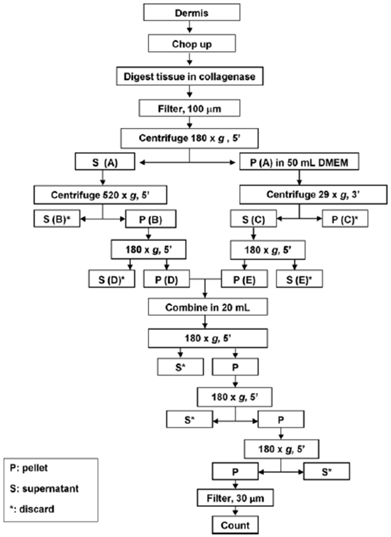
The hair follicle (HF) is a dynamic structure readily accessible within the skin, and contains various pools of stem cells that have a broad regenerative potential during normal homeostasis and in response to injury. Recent discoveries demonstrating the multipotent capabilities of hair follicle stem cells and the easy access to skin tissue make the HF an attractive source for isolating stem cells and their subsequent application in tissue engineering and regenerative medicine. Here, we describe the isolation and purification of hair follicle bulge stem cells from mouse skin, and hair reconstitution assays that allows the functional analysis of multipotent stem cells.
Keywords: Bulge; Hair follicle stem cell; Hair reconstitution assay; Regeneration.
Figures
Similar articles
-
Hair follicle: a novel source of multipotent stem cells for tissue engineering and regenerative medicine.Tissue Eng Part B Rev. 2013 Aug;19(4):265-78. doi: 10.1089/ten.TEB.2012.0422. Epub 2013 Jan 3. Tissue Eng Part B Rev. 2013. PMID: 23157470 Free PMC article.
-
Large-scale expansion and characterization of human adult neural crest-derived multipotent stem cells from hair follicle for regenerative medicine applications.Exp Oncol. 2017 Sep;39(3):171-180. Exp Oncol. 2017. PMID: 28967641
-
Multipotent nestin-positive, keratin-negative hair-follicle bulge stem cells can form neurons.Proc Natl Acad Sci U S A. 2005 Apr 12;102(15):5530-4. doi: 10.1073/pnas.0501263102. Epub 2005 Mar 31. Proc Natl Acad Sci U S A. 2005. PMID: 15802470 Free PMC article.
-
Hair follicle bulge: a fascinating reservoir of epithelial stem cells.J Dermatol Sci. 2007 May;46(2):81-9. doi: 10.1016/j.jdermsci.2006.12.002. Epub 2007 Jan 5. J Dermatol Sci. 2007. PMID: 17207970 Review.
-
Epithelial stem cells: a folliculocentric view.J Invest Dermatol. 2006 Jul;126(7):1459-68. doi: 10.1038/sj.jid.5700376. J Invest Dermatol. 2006. PMID: 16778814 Review.
Cited by
-
Hair Regeneration Methods Using Cells Derived from Human Hair Follicles and Challenges to Overcome.Cells. 2024 Dec 25;14(1):7. doi: 10.3390/cells14010007. Cells. 2024. PMID: 39791708 Free PMC article. Review.
-
Differential modularity of the mammalian Engrailed 1 enhancer network directs sweat gland development.PLoS Genet. 2023 Feb 6;19(2):e1010614. doi: 10.1371/journal.pgen.1010614. eCollection 2023 Feb. PLoS Genet. 2023. PMID: 36745673 Free PMC article.
-
Sulforaphane-Rich Broccoli Sprout Extract Promotes Hair Regrowth in an Androgenetic Alopecia Mouse Model via Enhanced Dihydrotestosterone Metabolism.Int J Mol Sci. 2025 Aug 1;26(15):7467. doi: 10.3390/ijms26157467. Int J Mol Sci. 2025. PMID: 40806594 Free PMC article.
-
An Ovol2-Zeb1 transcriptional circuit regulates epithelial directional migration and proliferation.EMBO Rep. 2019 Jan;20(1):e46273. doi: 10.15252/embr.201846273. Epub 2018 Nov 9. EMBO Rep. 2019. PMID: 30413481 Free PMC article.
-
Wnt signaling modulates mechanotransduction in the epidermis to drive hair follicle regeneration.Sci Adv. 2025 Feb 21;11(8):eadq0638. doi: 10.1126/sciadv.adq0638. Epub 2025 Feb 19. Sci Adv. 2025. PMID: 39970220 Free PMC article.
References
-
- Morris RJ, Liu Y, Marles L et al. (2004) Capturing and profiling adult hair follicle stem cells. Nat Biotechnol 22(4):411–417 - PubMed
-
- Cotsarelis G, Sun TT, Lavker RM (1990) Label-retaining cells reside in the bulge area of pilosebaceous unit: implications for follicular stem cells, hair cycle, and skin carcinogenesis. Cell 61(7):1329–1337 - PubMed
-
- Liu Y, Lyle S, Yang Z et al. (2003) Keratin 15 promoter targets putative epithelial stem cells in the hair follicle bulge. J Invest Dermatol 121(5):963–968 - PubMed
-
- Trempus CS, Morris RJ, Bortner CD et al. (2003) Enrichment for living murine keratinocytes from the hair follicle bulge with the cell surface marker CD34. J Invest Dermatol 120(4):501–511 - PubMed
-
- Morris RJ, Fischer SM, Klein-Szanto AJ et al. (1990) Subpopulations of primary adult murine epidermal basal cells sedimented on density gradients. Cell Tissue Kinet 23: 587–602 - PubMed
MeSH terms
Substances
Grants and funding
LinkOut - more resources
Full Text Sources
Other Literature Sources
Research Materials
Miscellaneous
