. 2016 Dec 1;44(21):10437-10453.
doi: 10.1093/nar/gkw649. Epub 2016 Jul 18.

Perlman syndrome nuclease DIS3L2 controls cytoplasmic non-coding RNAs and provides surveillance pathway for maturing snRNAs

Affiliations

Perlman syndrome nuclease DIS3L2 controls cytoplasmic non-coding RNAs and provides surveillance pathway for maturing snRNAs

Anna Łabno et al. Nucleic Acids Res. .

Abstract

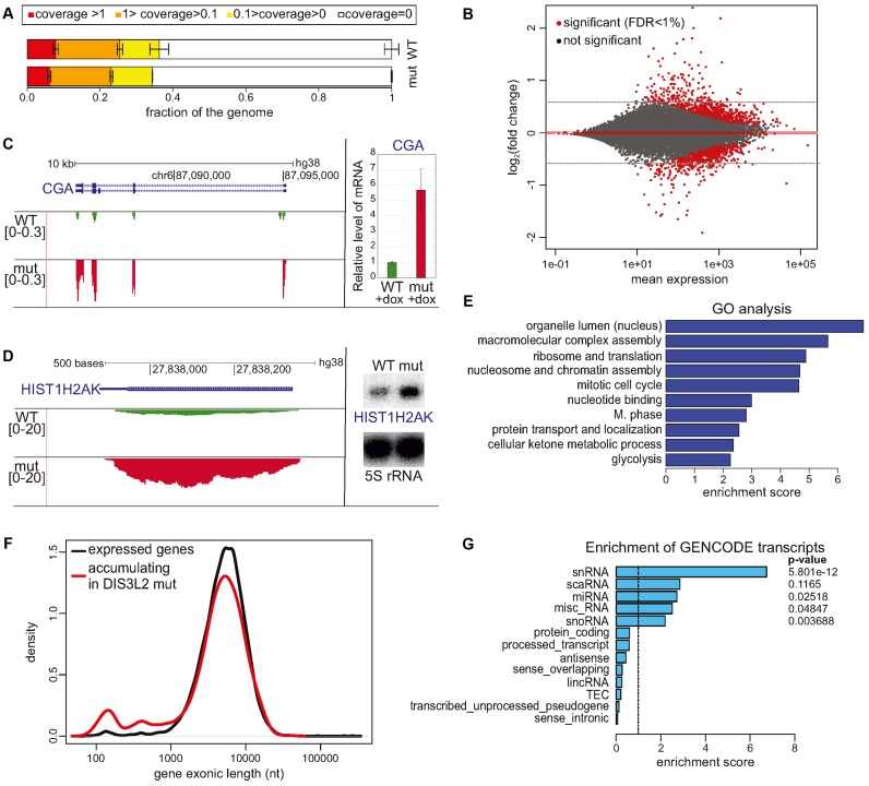
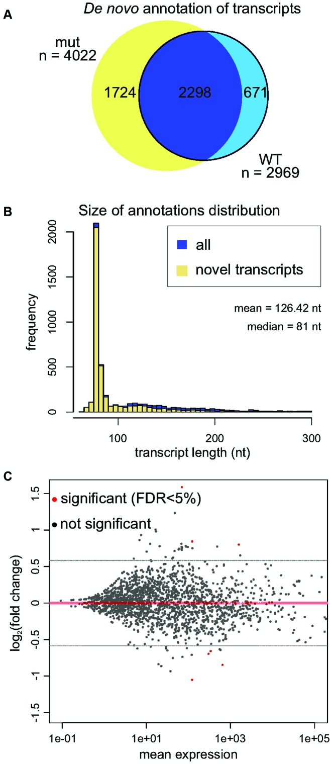
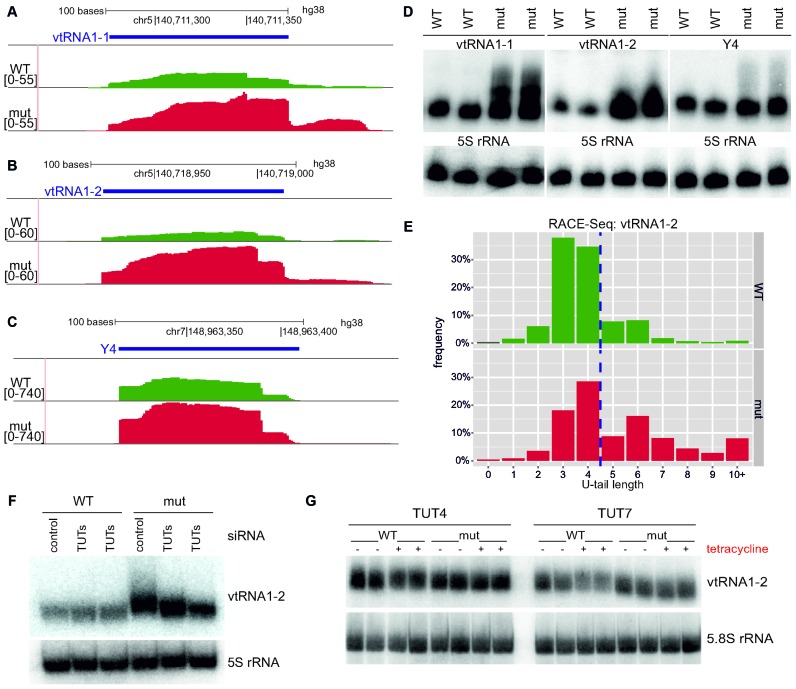
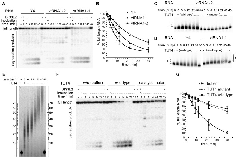
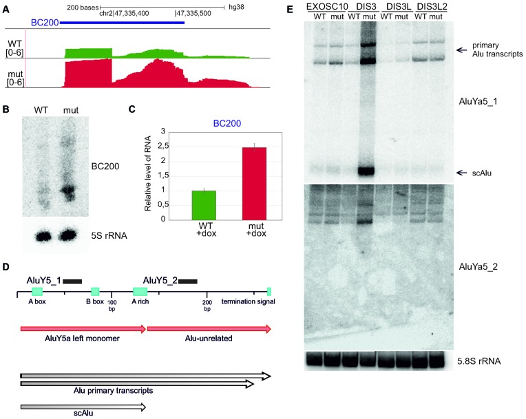
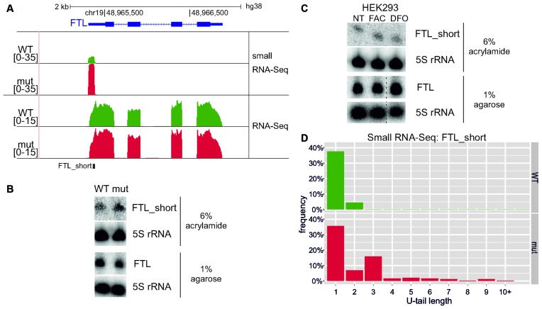
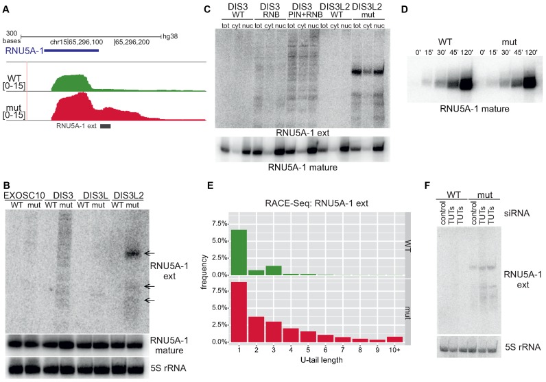
The exosome-independent exoribonuclease DIS3L2 is mutated in Perlman syndrome. Here, we used extensive global transcriptomic and targeted biochemical analyses to identify novel DIS3L2 substrates in human cells. We show that DIS3L2 regulates pol II transcripts, comprising selected canonical and histone-coding mRNAs, and a novel FTL_short RNA from the ferritin mRNA 5' UTR. Importantly, DIS3L2 contributes to surveillance of maturing snRNAs during their cytoplasmic processing. Among pol III transcripts, DIS3L2 particularly targets vault and Y RNAs and an Alu-like element BC200 RNA, but not Alu repeats, which are removed by exosome-associated DIS3. Using 3' RACE-Seq, we demonstrate that all novel DIS3L2 substrates are uridylated in vivo by TUT4/TUT7 poly(U) polymerases. Uridylation-dependent DIS3L2-mediated decay can be recapitulated in vitro, thus reinforcing the tight cooperation between DIS3L2 and TUTases. Together these results indicate that catalytically inactive DIS3L2, characteristic of Perlman syndrome, can lead to deregulation of its target RNAs to disturb transcriptome homeostasis.


Figures

Figure 1.
Figure 1.
Global analysis of DIS3L2 substrates. (A) RNA-Seq showed no significant difference between the genome fractions covered by RNA-Seq reads to given depths in WT and mutant DIS3L2 expressing cells. Bars represent the standard deviation for three biological replicates. (B) MAplot of total RNA sequencing results representing differential expression of DIS3L2 WT compared to DIS3L2 mut. Statistically significant hits (FDR < 1%) are color-coded red. Transcripts accumulating in the DIS3L2 mut have a positive value on the y-axis. (C and D) mRNAs are DIS3L2 substrates. Screenshots from Genome Browser showing genomic regions encoding CGA mRNA (C) or HIST1H2AK mRNA (D), with reads from deep sequencing for WT and mut DIS3L2. Number denotes the normalized expression measured by RNA-Seq (left); validation of CGA mRNA (C) and HIST1H2AK mRNA (D) accumulation in mut DIS3L2 by qPCR (bars represent the standard deviation for three biological replicates) and northern-blot (5S rRNA was used as an internal control), respectively (right). (E) Clustered GO analysis of genes accumulating in the DIS3L2 mutant. (F) Exon size distribution in genes that were upregulated in mutant DIS3L2 relative to all expressed genes is skewed towards a minor gene population with exons shorter than 1 kb and a maximum of approximately 150 nt. (G) Enrichment analysis of GENCODE transcript types among transcripts accumulating in DIS3L2 mutant cells compared to all expressed genes reveals a significant overrepresentation of snRNA. Other classes of short and non-protein coding RNA molecules, including miRNA, miscRNA and snoRNA, also showed moderate enrichment. Right, Fisher exact test P-value.
Figure 2.
Figure 2.
Analysis of short (20-200 nt) RNA DIS3L2 substrates. (A) Venn diagram summarizing de novo annotation of transcripts in the DIS3L2 mutant and WT samples. (B) Histogram representing distribution of size annotations in Cufflinks de novo transcriptome assembly. Potentially novel transcripts are shown in yellow. (C) MAplot of small RNA sequencing results demonstrates differential expression in DIS3L2 WT compared to DIS3L2 mutant. Statistically significant hits (FDR < 5%) are color-coded red. Transcripts accumulating in the DIS3L2 mut have a positive value on the y-axis.
Figure 3.
Figure 3.
Vault RNAs and Y RNAs are DIS3L2 substrates after TUT4/TUT7 uridylation (A–C). Screenshot from Genome Browser showing genomic region encoding vtRNA1-1 (A), vtRNA1-2 (B) and Y4 RNA (C), with reads from deep sequencing for WT and mut DIS3L2. Number denotes the normalized expression measured by RNA-Seq (left); (D) Validation of transcript accumulations shown in panels a-c in DIS3L2 mut as assessed by northern blot. 5S rRNA was used as an internal control. (E) RACE-Seq results showing that vtRNA1-2 transcripts bearing 3′ non-templated nucleotide additions accumulated in cells expressing catalytically inactive DIS3L2. Since vtRNA1-2 is a polymerase III transcript, it has four uridines encoded in the genome (left side of dashed line). The distribution of reads with the selected U-tail length is shown (bars represent frequency of reads with selected U-tail length normalized to total read counts). (F) Northern blot analysis of vtRNA1-2 in WT and mut DIS3L2 cell lines after siRNA-mediated TUTase repression. 5S rRNA was used as an internal control. (G) Northern blot analysis of vtRNA1-2 RNA in HEK293 parental cell line after transfection with plasmids for tetracycline-inducible overexpression of wild type or catalytically inactive TUT4 or TUT7. ‘+’ indicates tetracycline induction and ‘-’ represents no induction. 5.8S rRNA was used as an internal control.
Figure 4.
Figure 4.
DIS3L2 preferentially degrades TUTase-uridylated substrates in vitro. (A) A typical time-course experiment visualized by denaturing PAGE and phosphorimaging. In vitro transcribed, 5′ radioactively labelled Y4, vtRNA1-2 and vtRNA1-1 were incubated with purified recombinant DIS3L2 protein. Aliquots were withdrawn at the indicated time points and reactions were stopped rapidly. (B) Results of 6 independent time-course experiments were quantified by Multi Gauge software, plotted and fit with a single exponential decay equation using Graph Pad Prism software. (C) In vitro transcribed, 5′ radioactively labelled vtRNA1-2 was incubated in the presence of either wild type or catalytically inactive (D1011A) TUT4. Reactions were rapidly stopped, separated by 10% sequencing PAGE and visualized by phosphorimaging. The ladder on the left indicates the number of uridines added to the 3′ end of the RNA. (D) As in panel C with Y4 and vtRNA1-1 used as substrates in the presence of WT TUT4. (E) As in panels C and D but a 5′ fluorescently labelled (FAM) 44 mer RNA oligonucleotide was the substrate. The RNAs were separated by 6% PAGE and fluorescent signals were visualized with a Typhoon FLA-9000 imaging scanner (GE Healthcare). (F) Representative PAGE analysis of a combined extension-degradation time-course assay of vtRNA1-2 in the presence of DIS3L2 and either wild-type or catalytically inactive mutant TUT4 or buffer (control w/o TUT). The assay was visualized as in panel A. (G) Plot summarizing results of four independent extension-degradation time-course assays (as in panel F). The graph was generated as in panel B.
Figure 5.
Figure 5.
BC200, an Alu-like element RNA, is a DIS3L2 substrate, while DIS3 degrades transcripts from Alu-repeats. (A) Screenshot from Genome Browser showing the genomic region encoding BC200 RNA with reads from deep sequencing for WT and mut DIS3L2. Number denotes the normalized expression measured by RNA-Seq. (B) Validation of BC200 RNA accumulation in DIS3L2 mut by northern blot. 5S rRNA was used as an internal control. (C) Validation of BC200 accumulation in mut DIS3L2 by qPCR. Bars represent the standard deviation for three biological replicates. (D) Example of transcripts arising from Alu repeats (AluYa5) accumulating in DIS3 mut, but not in EXOSC10, DIS3L or DIS3L2 mutants. Schematic representation of the AluYa5 transcript. Northern blot probe binding sites are shown for AluY5_1 and AluY5_2. (E) Northern blot showing accumulation of AluYa5 transcripts in DIS3 mut cells, including scAlu (small cytoplasmic Alu) (produced from AluY5a left monomer). 5.8S rRNA was used as an internal control.
Figure 6.
Figure 6.
Identification of FTL_short, a novel transcript from the 5′-UTR of ferritin mRNA. (A) Screenshot from Genome Browser showing genomic region encoding FTL mRNA with RNA-Seq and small RNA-Seq reads (see Materials and Methods) for WT and mut DIS3L2 cells. Number denotes the normalized expression measured by RNA-Seq. (B) Validation of FTL_short by northern blot after 6% PAGE. The amount of full-length ferritin mRNA remained constant, as shown by northern blot after electrophoresis in 1% agarose. 5S rRNA was used as an internal control for both cases. (C) FTL_short possibly does not function in iron metabolism. Parental HEK293 cells were treated for 24 h with substances that alter environmental iron levels: FAC (ferric ammonium citrate) and DFO (deferoxamine). The amounts of FTL_short and full-length ferritin transcript were then analysed by northern blot. 5S rRNA was used as an internal control in both cases. Dashed lines indicate degraded RNA samples that were excised (second biological replicate). (D) RNA-Seq results showing that FTL_short transcripts bearing 3′ non-templated nucleotide additions accumulated in cells expressing catalytically inactive DIS3L2. The distribution of reads with the selected U-tail length is shown (bars represent frequency of 3′ end reads with selected U-tail length normalized to the total number of reads maping to 3′ end).
Figure 7.
Figure 7.
DIS3L2-mediated surveillance of maturing U5 snRNA. (A) Screenshot from Genome Browser showing genomic region encoding U5 snRNA with deep sequencing reads for WT and mut DIS3L2. Number denotes the normalized expression measured by RNA-Seq. (B) Northern blot verification of extended snRNAs (‘ext’) in model cell lines producing wild-type or mutated EXOSC10, DIS3, DIS3L or DIS3L2. The level of mature snRNAs was also analyzed. 5S rRNA was used as an internal control. (C) Extended snRNAs accumulating in the presence of mutated DIS3 and DIS3L2 are present in the cytoplasm and the nucleus. The localization of extended snRNAs in different cellular compartments was assessed by cell fractionation and northern blot analysis (tot – total RNA, nuc – nuclear RNA, cyt – cytoplasmic RNA). Analysis of mature U5 snRNA localisation after cell fractionation was also conducted. Control of cell fractionation is identical as in Supplementary Figure S7. (D) Accumulation of extended U5 snRNAs does not influence production of mature snRNA species. 4-thiouridine (4sU) labelling was used to study the kinetics of mature snRNA production. RNA was collected after labelling and terminating reactions at the indicated time points. RNA labelled with 4sU was retrieved by biotinylation and separation on streptavidin beads. (E) Extended U5 snRNA transcripts bearing 3′ non-templated nucleotide additions accumulate in cells expressing catalytically inactive DIS3L2 as shown by RACE-Seq. The distribution of reads with selected U-tail lengths is shown (bars represent the frequency of reads with selected U-tail length normalized to total read counts). (F) Uridylation enhances degradation of extended snRNAs. TUT4 and TUT7 were silenced in model cell lines using siRNA (lanes TUTs) and the presence of extended snRNA transcripts was monitored by northern blot. 5S rRNA was used as an internal control.

Similar articles

Cited by

References

    1. Mroczek S., Kufel J. Apoptotic signals induce specific degradation of ribosomal RNA in yeast. Nucleic Acids Res. 2008;36:2874–2888. - PMC - PubMed
    1. Siwaszek A., Ukleja M., Dziembowski A. Proteins involved in the degradation of cytoplasmic mRNA in the major eukaryotic model systems. RNA Biol. 2014;11:1122–1136. - PMC - PubMed
    1. Kung J.T.Y., Colognori D., Lee J.T. Long noncoding RNAs: past, present, and future. Genetics. 2013;193:651–669. - PMC - PubMed
    1. Fu X.-D. Non-coding RNA: a new frontier in regulatory biology. Natl. Sci. Rev. 2014;1:190–204. - PMC - PubMed
    1. Tomecki R., Kristiansen M.S., Lykke-Andersen S., Chlebowski A., Larsen K.M., Szczesny R.J., Drazkowska K., Pastula A., Andersen J.S., Stepien P.P., et al. The human core exosome interacts with differentially localized processive RNases: hDIS3 and hDIS3L. EMBO J. 2010;29:2342–2357. - PMC - PubMed

MeSH terms

Supplementary concepts