Review
. 2016 Jul 17:36:571-602.
doi: 10.1146/annurev-nutr-071715-051110.

Lutein and Zeaxanthin Isomers in Eye Health and Disease

Affiliations
Review

Lutein and Zeaxanthin Isomers in Eye Health and Disease

Julie Mares. Annu Rev Nutr. .

Abstract

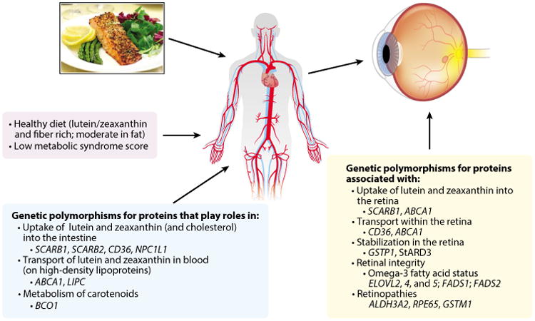
Current evidence suggests lutein and its isomers play important roles in ocular development in utero and throughout the life span, in vision performance in young and later adulthood, and in lowering risk for the development of common age-related eye diseases in older age. These xanthophyll (oxygen-containing) carotenoids are found in a wide variety of vegetables and fruits, and they are present in especially high concentrations in leafy green vegetables. Additionally, egg yolks and human milk appear to be bioavailable sources. The prevalence of lutein, zeaxanthin, and meso-zeaxanthin in supplements is increasing. Setting optimal and safe ranges of intake requires additional research, particularly in pregnant and lactating women. Accumulating evidence about variable interindividual response to dietary intake of these carotenoids, based on genetic or metabolic influences, suggests that there may be subgroups that benefit from higher levels of intake and/or alternate strategies to improve lutein and zeaxanthin status.

Keywords: carotenoids; cataract; macula; macular degeneration; retinopathy; vision.


Figures

Figure 1
Figure 1
Image of the human eye.
Figure 2
Figure 2
Cross-section of a primate retina, in the macula, photographed in either white or blue light, indicating macular pigment (composed of lutein, zeaxanthin, and meso-zeaxanthin) in retinal layers and its absorption of blue light from macular pigment. Figure adapted with permission from the American Journal of Clinical Nutrition and D. Max Snodderly (170).
Figure 3
Figure 3
The concentration of macular pigment (MP) in human eyes and ratio of lutein (L) to zeaxanthin (Z) in relation to distance from the center of the fovea within the macula. Figure courtesy of John Landrum and Richard Bone, as published in Carotenoids and Retinal Disease, CRC Press (98). Adapted with permission.
Figure 4
Figure 4
Distribution of proteins affecting or affected by macular xanthophylls in primate retina. Full names for genes are available from http://www.ncbi.nlm.nih.gov/gene. Superscripts on gene symbols refer to reference numbers of immunolocalization studies containing micrographs in Reference 161. Abbreviations: Am, amacrine cell; B, bipolar cell; BrM, Bruch's membrane; C, cone photoreceptors; CC, retinal choroid layer; DA, displaced amacrine cell; ELM, external limiting membrane; G, ganglion cell; GCL, ganglion cell layer; H, horizontal cell; ILM, inner limiting membrane; INL, inner nuclear layer; IPL, inner plexiform layer (interneurons); M, Muller cell; NFL, nerve fiber layer; ONL, outer nuclear layer; OPL, outer plexiform layer; PRIS, photoreceptor inner segment; PROS, photoreceptor outer segment; R, rod photoreceptor; RPEa, retinal pigment epithelium apical area; RPEb, retinal pigment epithelium basal area. Retinal layers with macular xanthophyll concentrations. Schematic created by D. Fisher and adapted with permission from the American Journal of Clinical Nutrition (161) and John Paul SanGiovanni.
Figure 5
Figure 5
Phenotypes and genotypes associated with macular pigment optical density in the Carotenoids in Age-Related Eye Disease Study (120, 126, 128, 129). The evidence suggests that the indicated aspects of diet, metabolism, and genotype might influence the absorption and transport of lutein and zeaxanthin in the blood (left and center boxes) and that the indicated genotypes and activities of the proteins these genes encode (right box) might influence their uptake and/or stabilization in the retina.

References

    1. Age-Related Eye Disease Study Investig. Lutein + zeaxanthin and omega-3 fatty acids for age-related macular degeneration: the Age-Related Eye Disease Study 2 (AREDS2) randomized clinical trial. JAMA. 2013;309:2005–15. - PubMed
    1. Akuffo KO, Nolan JM, Howard AN, Moran R, Stack J, et al. Sustained supplementation and monitored response with differing carotenoid formulations in early age-related macular degeneration. Eye (Lond) 2015;29:902–12. - PMC - PubMed
    1. Aleman TS, Cideciyan AV, Windsor EA, Schwartz SB, Swider M, et al. Macular pigment and lutein supplementation in ABCA4-associated retinal degenerations. Investig Ophthalmol Vis Sci. 2007;48:1319–29. - PMC - PubMed
    1. Aleman TS, Duncan JL, Bieber ML, de Castro E, Marks DA, et al. Macular pigment and lutein supplementation in retinitis pigmentosa and Usher syndrome. Investig Ophthalmol Vis Sci. 2001;42:1873–81. - PubMed
    1. Amengual J, Lobo GP, Golczak M, Li HN, Klimova T, et al. A mitochondrial enzyme degrades carotenoids and protects against oxidative stress. FASEB J. 2011;25:948–59. - PMC - PubMed

MeSH terms