. 2016 Nov;1860(11 Pt A):2428-2437.
doi: 10.1016/j.bbagen.2016.07.010. Epub 2016 Jul 16.

Nitro-oleic acid inhibits vascular endothelial inflammatory responses and the endothelial-mesenchymal transition

Affiliations

Nitro-oleic acid inhibits vascular endothelial inflammatory responses and the endothelial-mesenchymal transition

Gabriela Ambrozova et al. Biochim Biophys Acta. 2016 Nov.

Abstract

Background: Inflammatory-mediated pathological processes in the endothelium arise as a consequence of the dysregulation of vascular homeostasis. Of particular importance are mediators produced by stimulated monocytes/macrophages inducing activation of endothelial cells (ECs). This is manifested by excessive soluble pro-inflammatory mediator production and cell surface adhesion molecule expression. Nitro-fatty acids are endogenous products of metabolic and inflammatory reactions that display immuno-regulatory potential and may represent a novel therapeutic strategy to treat inflammatory diseases. The purpose of our study was to characterize the effects of nitro-oleic acid (OA-NO2) on inflammatory responses and the endothelial-mesenchymal transition (EndMT) in ECs that is a consequence of the altered healing phase of the immune response.

Methods: The effect of OA-NO2 on inflammatory responses and EndMT was determined in murine macrophages and murine and human ECs using Western blotting, ELISA, immunostaining, and functional assays.

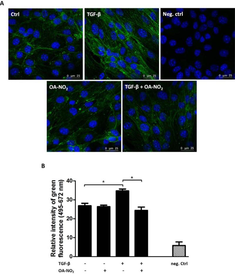
Results: OA-NO2 limited the activation of macrophages and ECs by reducing pro-inflammatory cytokine production and adhesion molecule expression through its modulation of STAT, MAPK and NF-κB-regulated signaling. OA-NO2 also decreased transforming growth factor-β-stimulated EndMT and pro-fibrotic phenotype of ECs. These effects are related to the downregulation of Smad2/3.

Conclusions: The study shows the pleiotropic effect of OA-NO2 on regulating EC-macrophage interactions during the immune response and suggests a role for OA-NO2 in the regulation of vascular endothelial immune and fibrotic responses arising during chronic inflammation.

General significance: These findings propose the OA-NO2 may be useful as a novel therapeutic agent for treatment of cardiovascular disorders associated with dysregulation of the endothelial immune response.

Keywords: Endothelial cells; Endothelial-mesenchymal transition; Macrophages; Nitro-fatty acids; Nitro-oleic acid; Vascular inflammation.


Figures

Fig. 1
Fig. 1. OA-NO2 regulates cytokine production in activated ECs
MS-1 cells were treated with or without OA-NO2 (1.0 μM) and the following cytokines for 24 h: IFN-γ (50 ng/ml), IL-1β (5 ng/ml), and TNF-α (10 ng/ml). RANTES (A-C), IL-6 (D-F), MCP-5 (G), and GM-CSF (H) levels were detected by ELISA. The data represent 3-6 independent experiments (each determined in duplicate). A *p value of less than 0.05 was considered significant when evaluating differences between the individual bars.
Fig. 2
Fig. 2. OA-NO2 decreases IFN-γ-induced ICAM-1 expression and adhesive properties in ECs
MS-1 cells were treated with or without OA-NO2 (1.0 μM) and IFN-γ (50 ng/ml) for 24 h. ICAM-1 expression (green) was detected by immunostaining (A), nuclei were stained with DAPI (blue) [45]. Typical representative figures are shown from n=3 independent replicates. The expression of ICAM-1 was detected also by WB (B). Adhesion of Calcein AM-stained RAW 264.7 macrophages to OA-NO2 (1.0 μM)- and IFN-γ (50 ng/ml)-treated MS-1 ECs was observed after 1 h co-incubation (C). Relative fluorescence intensity of Calcein AM was measured (ex. 480, em. 530 nm, determined in duplicates). The data represent the ratios between individual values for the optical densities of bands determined for ICAM-1 and housekeeping protein (WB) or means from fluorescence measurements (adherence assay), n=3-4. A *p value of less than 0.05 was considered significant when evaluating differences between the individual bars.
Fig. 3
Fig. 3. Signaling pathways affected by OA-NO2 in activated MS-1 ECs
MS-1 cells were treated with or without OA-NO2 (1.0 μM) and selected inflammatory mediators IFN-γ (50 ng/ml), IL-1β (5 ng/ml), TNF-α (10 ng/ml), and TGF-β (10 ng/ml). STAT1 (A) and STAT3 (B) phosphorylation was detected after 30 min, MAPK (C-E) and NF-κB (F) phosphorylation after 20 min. The data represent the ratios between individual values for the optical densities of bands determined for the phosphorylated and total forms of each protein (n=3-5). A *p value of less than 0.05 was considered significant when evaluating differences between the individual bars.
Fig. 4
Fig. 4. OA-NO2 regulates TGF-β-triggered changes in actin cytoskeleton in ECs
MS-1 cells were treated with or without OA-NO2 (1.0 μM) and TGF-β (10 ng/ml) for 48 h. Immunostaining was used to identify filamentous actin stress fibers (green) characteristic of active fibroblasts (A). Nuclei were stained with DAPI (blue) [45]. Typical representative figures are shown from n=3-6 independent replicates. The mean value of the optical intensity of the green component (ex. 495, em. 672 nm) of each image was calculated (B). A *p value of less than 0.05 was considered significant when evaluating differences between the individual bars.
Fig. 5
Fig. 5. OA-NO2 decreases the TGF-β-induced FSP1 and α-SMA expression in ECs
MS-1 cells were treated with or without OA-NO2 (1.0 μM) and TGF-β (10 ng/ml). The expression of fibroblast marker FSP-1 (green) was detected by immunostaining after 48 h of treatment (A). Nuclei were stained with DAPI (blue) [45]. Typical representative figures are shown from n=3 independent replicates. The mean value of the optical intensity of the green component (ex. 495, em. 672 nm) of each image was calculated (B). The expression of α-SMA was determined in MS-1 cells treated for 6 d by WB (C). The data represent the ratios between individual values for the optical densities of bands determined for α-SMA and housekeeping protein (n=4). A *p value of less than 0.05 was considered significant when evaluating differences between the individual bars.
Fig. 6
Fig. 6. OA-NO2 decreases the TGF-β-induced Smad2/3 activation in ECs
MS-1 cells were treated with or without OA-NO2 (1.0 μM) and TGF-β (10 ng/ml). The expressions of p-Smad2/t-Smad2 (A) and p-Smad3/t-Smad3 (B) were detected after 30 min of treatment. The data displayed in graphs (n=3-5) represent the ratios between individual values for the optical densities of bands determined for the phosphorylated and total forms of each protein. A *p value of less than 0.05 was considered significant when evaluating differences between the individual bars.

Similar articles

Cited by

References

    1. Popa C, Netea MG, van Riel PL, van der Meer JW, Stalenhoef AF. The role of TNF-alpha in chronic inflammatory conditions, intermediary metabolism, and cardiovascular risk. Journal of lipid research. 2007;48:751–762. - PubMed
    1. Ambrozova G, Martiskova H, Koudelka A, Ravekes T, Rudolph TK, Klinke A, Rudolph V, Freeman BA, Woodcock SR, Kubala L, Pekarova M. Nitro-oleic acid modulates classical and regulatory activation of macrophages and their involvement in pro-fibrotic responses. Free radical biology & medicine. 2015 - PMC - PubMed
    1. Khoo NK, Rudolph V, Cole MP, Golin-Bisello F, Schopfer FJ, Woodcock SR, Batthyany C, Freeman BA. Activation of vascular endothelial nitric oxide synthase and heme oxygenase-1 expression by electrophilic nitro-fatty acids. Free radical biology & medicine. 2010;48:230–239. - PMC - PubMed
    1. Mai J, Virtue A, Shen J, Wang H, Yang XF. An evolving new paradigm: endothelial cells--conditional innate immune cells. Journal of hematology & oncology. 2013;6:61. - PMC - PubMed
    1. Lawson C, Wolf S. ICAM-1 signaling in endothelial cells. Pharmacological reports : PR. 2009;61:22–32. - PubMed

Publication types

MeSH terms

LinkOut - more resources