. 2016 Jul 19:16:496.
doi: 10.1186/s12885-016-2521-9.

New anti-cancer chemicals Ertredin and its derivatives, regulate oxidative phosphorylation and glycolysis and suppress sphere formation in vitro and tumor growth in EGFRvIII-transformed cells

Affiliations

New anti-cancer chemicals Ertredin and its derivatives, regulate oxidative phosphorylation and glycolysis and suppress sphere formation in vitro and tumor growth in EGFRvIII-transformed cells

Sonoko Atsumi et al. BMC Cancer. .

Abstract

Background: EGFRvIII is a mutant form of the epidermal growth factor receptor gene (EGFR) that lacks exons 2-7. The resulting protein does not bind to ligands and is constitutively activated. The expression of EGFRvIII is likely confined to various types of cancer, particularly glioblastomas. Although an anti-EGFRvIII vaccine is of great interest, low-molecular-weight substances are needed to obtain better therapeutic efficacy. Thus, the purpose of this study is to identify low molecular weight substances that can suppress EGFRvIII-dependent transformation.

Methods: We constructed a new throughput screening system and searched for substances that decreased cell survival of NIH3T3/EGFRvIII spheres under 3-dimensional (3D)-culture conditions, but retained normal NIH3T3 cell growth under 2D-culture conditions. In vivo activity was examined using a mouse transplantation model, and derivatives were chemically synthesized. Functional characterization of the candidate molecules was investigated using an EGFR kinase assay, immunoprecipitation, western blotting, microarray analysis, quantitative polymerase chain reaction analysis, and measurement of lactate and ATP synthesis.

Results: In the course of screening 30,000 substances, a reagent, "Ertredin" was found to inhibit anchorage-independent 3D growth of sphere-forming cells transfected with EGFRvIII cDNA. Ertredin also inhibited sphere formation in cells expressing wild-type EGFR in the presence of EGF. However, it did not affect anchorage-dependent 2D growth of parental NIH3T3 cells. The 3D-growth-inhibitory activity of some derivatives, including those with new structures, was similar to Ertredin. Furthermore, we demonstrated that Ertredin suppressed tumor growth in an allograft transplantation mouse model injected with EGFRvIII- or wild-type EGFR-expressing cells; a clear toxicity to host animals was not observed. Functional characterization of Ertredin in cells expressing EGFRvIII indicated that it stimulated EGFRvIII ubiquitination, suppressed both oxidative phosphorylation and glycolysis under 3D conditions, and promoted cell apoptosis.

Conclusion: We developed a high throughput screening method based on anchorage-independent sphere formation induced by EGFRvIII-dependent transformation. In the course of screening, we identified Ertredin, which inhibited anchorage-independent 3D growth and tumor formation in nude mice. Functional analysis suggests that Ertredin suppresses both mitochondrial oxidative phosphorylation and cytosolic glycolysis in addition to promoting EGFRvIII degradation, and stimulates apoptosis in sphere-forming, EGFRvIII-overexpressing cells.

Keywords: 3D; Anchorage-independent; Apoptosis; EGFRvIII; Ertredin; Glycolysis; Oxidative phosphorylation; Sphere.


Figures
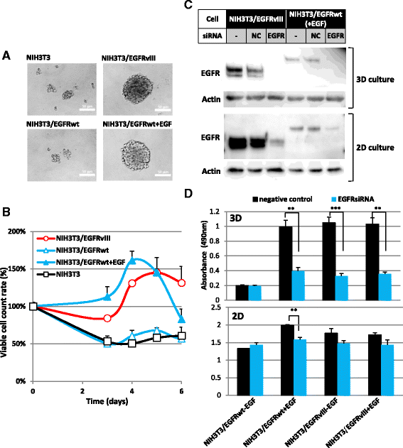
Fig. 1
Fig. 1
Overexpression of EGFRvIII or EGFRwt induced anchorage-independent growth of NIH3T3 cells. a Anchorage-independent (3D) growth of NIH3T3/EGFRvIII or NIH3T3/EGFRwt cells. Cells (2 × 105 cells/mL) were seeded and observed by phase-contrast microscopy after cultivation for 3 days. Images were acquired using an Olympus DP71 microscope camera and processed by Adobe Photoshop. b Growth curves of NIH3T3 cells overexpressing EGFRvIII or EGFRwt in 3D cell cultures. Cells (1 × 105 cells/mL, 100 μl) were seeded on ULAS 96-well plates on day 0. Viable cells were counted at the indicated times by CellTiter-Glo Luminescent Cell Viability Assay. c EGFR-siRNA inhibited EGFRvIII and EGFRwt protein expression in NIH3T3 cells overexpressing each protein. Cells were lysed and 6 μg protein was applied to 10 % SDS-PAGE gel. Cells were cultured on ULAS plate for 3 days (3D) or on a normal cell-attachment plate for 1 day (2D) after EGFR-siRNA transfection. d EGFR-siRNA inhibited NIH3T3/EGFRvIII and NIH3T3/EGFRwt anchorage-independent 3D growth. Viable cell counts were measured by CellTiter 96 AQueous One Solution Cell Proliferation Assay 3 days after transfection with EGFR-siRNA (NM005228_stealth_2438). Cells were cultured with (50 ng/mL) or without EGF on ULAS plates (upper, 3D) or on normal tissue culture plate (lower, 2D)
Fig. 2
Fig. 2
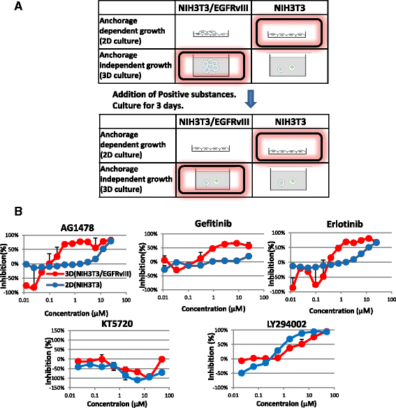
High throughput screening system developed to identify inhibitors of anchorage-independent growth induced by EGFRvIII. a Outline of the screening system. b Potency of protein kinase inhibitors in the screening systems. NIH3T3/EGFRvIII cells were seeded on ULAS 96-well plates for 3D-culture (red line). NIH3T3 cells were seeded on normal tissue culture plates for 2D-culture (blue line). Each inhibitor was added to the medium on day 0. Three days after inoculation, viable cells were counted and the extent of inhibition was calculated
Fig. 3
Fig. 3
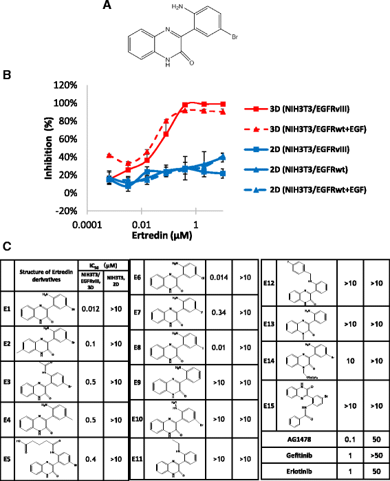
Ertredin derivatives inhibited anchorage-independent growth of NIH3T3/EGFRvIII cells. a Structure of Ertredin. b Inhibitory activity of Ertredin against 3D growth of NIH3T3/EGFRvIII (red solid line) or NIH3T3/EGFRwt (red dashed line), and 2D growth of NIH3T3. Cells were seeded and cultured on ULAS plates (3D) or normal tissue culture plates (2D). Extent of inhibition was detected on day 3. EGF (100 ng/mL) was added to NIH/3 T3/EGFRwt 3D culture. c Structure-activity relationships of Ertredin derivatives
Fig. 4
Fig. 4
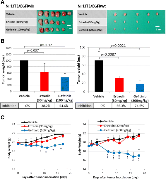
Ertredin suppressed NIH3T3/EGFRvIII (left) and NIH3T3/EGFRwt (right) tumorigenicity in nude mice. a Macroscopic tumor appearance at day 17 (NIH3T3/EGFRvIII) and day 18 (NIH3T3/EGFRwt). b Weights of tumors at day 17 (NIH3T3/EGFRvIII) and day 18 (NIH3T3/EGFRwt). c Time-dependent changes in body weights of mice transplanted with NIH3T3/EGFRvIII (left) and NIH3T3/EGFRwt. P values in two-tailed paired student t-test are shown: *p < 0.05; **p < 0.01; ***p <0.001
Fig. 5
Fig. 5
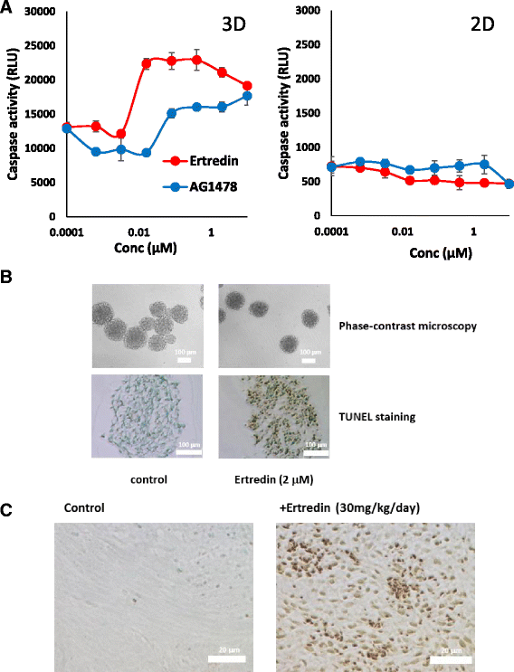
Ertredin induced apoptosis in vitro 3D-sphere and in vivo tumors of NIH3T3/EGFRvIII cells. a Cells with the addition of Ertredin (red line) or AG1478 (blue line) were seeded on ULAS plates (left) or tissue culture treated plates (right), followed by cultivation for 72 h. Caspase 3/7 activities were measured as described in Materials and Methods. b (Upper) Spheres were observed by phase contrast microscopy 72 h after seeding of cells on ULAS plates. (Lower) Apoptotic cells (brown) in paraffin sections were determined by TUNEL assay. Cells were cultured in medium with Ertredin (2 μM) (right) or vehicle (left). Apoptotic cells were stained brown. DNA counterstained with Methyl Green. c TUNEL staining for the detection of apoptotic cells in tumors isolated from mice treated with Ertredin (right) or control mice without Ertredin (left)
Fig. 6
Fig. 6
Ertredin did not directly inhibit EGFR kinase but stimulated ubiquitination of EGFR in NIH3T3/EGFRvIII cells. a Direct effect of Ertredin (red line) or AG1478 (blue line) on the tyrosine kinase of human EGFR. b Effect of Ertredin on autophosphorylation of EGFRvIII in NIH3T3/EGFRvIII cells. Cells (3 × 105 /mL, 5 mL) were seeded on ULAS 6-well plates and cultured for 22 h. Ertredin or AG1478 was then added to a final concentration of 1 μM. Cells were harvested at the indicated time after the addition of chemicals. Each cell lysate (16 μg of protein) was applied on 10 % SDS-PAGE followed by immunoblot analysis. c NIH3T3/EGFRvIII spheres in ULAS dishes were treated with Ertredin (final concentration, 2 μM), AG1478 (final concentration, 2 μM), or vehicle for 17 h in the presence of MG132 (50 μM). Lysates were prepared from the cells and subjected to immunoprecipitation with anti-EGFR antibody followed by western blot with respective antibodies as shown (lane 1–3). Input lanes indicate 3.7 % of lysate used in immunoprecipitation (lane 4–6). The triangles indicate the estimated position of EGFRvIII
Fig. 7
Fig. 7
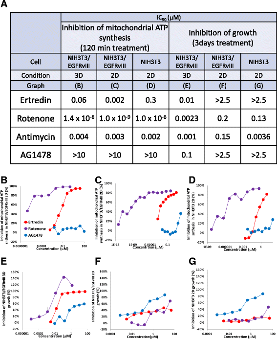
Ertredin inhibited mitochondrial ATP synthesis in NIH3T3/EGFRvIII cells under 3D conditions. a Summary of the assay results. Inhibition of mitochondrial ATP synthesis by 120 min treatment with Ertredin (red line), Rotenone (purple line), AG1478 (blue line) in b NIH3T3/EGFRvIII cells under 3D-culture conditions, c NIH3T3/EGFRvIII cells under 2D-culture conditions, and d NIH3T3 cells under 2D-culture conditions. The cells were cultured in absence of glucose (in galactose-containing medium) and then ATP synthesis was measured. Inhibition of cell growth with the addition of the substances for 3 days in e NIH3T3/EGFRvIII cells under 3D-culture conditions, f NIH3T3/EGFRvIII cells under 2D-culture conditions, and g NIH3T3 cells under 2D-culture conditions
Fig. 8
Fig. 8
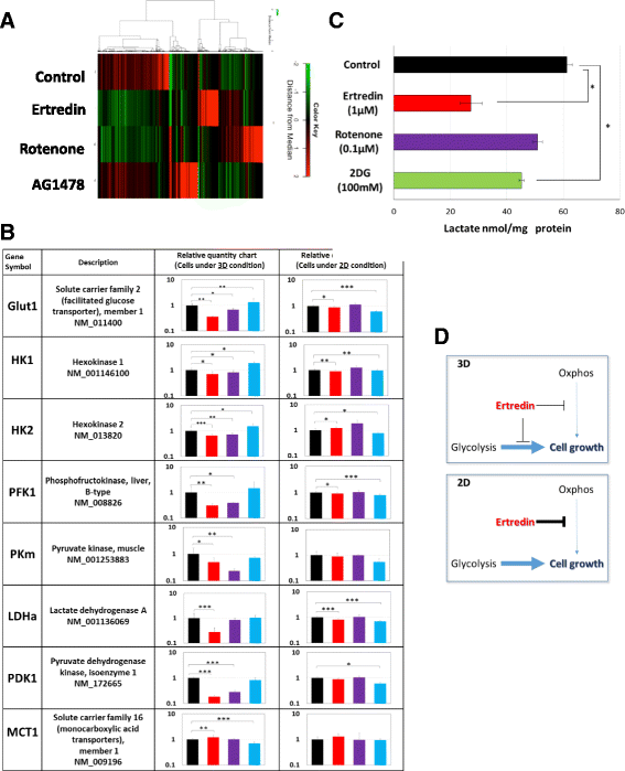
Ertredin suppresses the expression of glycolytic pathway enzymes in NIH/3 T3 EGFRvIII cells under 3D conditions. NIH3T3/EGFRvIII cells (2 × 106 cells/well) were seeded on a ULAS 6-well plate for 3D culture and 10 cm tissue culture-treated plate for 2D cell culture. Ertredin (2 μM), Rotenone (1.25 μM) AG1478 (2 μM) or DMSO (0.1 %) was added to the medium and after 24 h of culture, the cells were harvested and total RNA was isolated. a Microarray heat map. b Relative quantitative charts obtained by qPCR analysis for each glycolysis pathway gene in cells treated with Ertredin (red bar), Rotenone (purple bar), AG1478 (blue bar), and DMSO (black bar) under 3D and 2D conditions. c Lactate in the cells cultured with the indicated substances for 24 h under 3D conditions. 2-deoxyglucose (2DG), a glucose analogue, is known to inhibit hexokinase, the first step of glycolysis. d An explanatory diagram of cell-growth inhibition under 3D conditions by Ertredin

Similar articles

Cited by

References

    1. Sebastian S, Settleman J, Reshkin SJ, Azzariti A, Bellizzi A, Paradiso A. The complexity of targeting EGFR signaling in cancer: from expression to turnover. Biochim Biophys Acta. 2006;1766(1):120–139. - PubMed
    1. Congdon KL, Gedeon PC, Suryadevara CM, Caruso HG, Cooper LJ, Heimberger AB, Sampson JH. Epidermal growth factor receptor and variant III targeted immunotherapy. Neuro Oncol. 2014;16 Suppl 8:viii20–25. - PMC - PubMed
    1. Cohen AL, Colman H. Glioma biology and molecular markers. Cancer Treat Res. 2015;163:15–30. doi: 10.1007/978-3-319-12048-5_2. - DOI - PubMed
    1. Saito T, Mitomi H, Yao T. Molecular pathology and potential therapeutic targets in esophageal basaloid squamous cell carcinoma. Int J Clin Exp Pathol. 2015;8(3):2267–2273. - PMC - PubMed
    1. Bejar C, Maubec E. Therapy of advanced squamous cell carcinoma of the skin. Curr Treat Options Oncol. 2014;15(2):302–320. doi: 10.1007/s11864-014-0280-x. - DOI - PubMed

MeSH terms