. 2016 Jul 19:16:495.
doi: 10.1186/s12885-016-2525-5.

MicroRNA-183 suppresses cancer stem-like cell properties in EBV-associated nasopharyngeal carcinoma

Affiliations

MicroRNA-183 suppresses cancer stem-like cell properties in EBV-associated nasopharyngeal carcinoma

Chartia Ching-Mei Cheung et al. BMC Cancer. .

Abstract

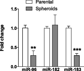
Background: Nasopharyngeal carcinoma (NPC) is an Epstein-Barr virus (EBV)-associated epithelial malignancy that exhibits distinct geographical and ethnic prevalence. Although the contemporary therapeutic approach of radio-/chemotherapy provides excellent results for patients with early-stage disease, it is far from satisfactory for those with disease remission and distant metastasis. Promising therapeutic strategies for advanced and relapsed NPC are still lacking. We recently identified and characterized a cancer stem-like cell (CSC) subpopulation in NPC that appeared to play an important role in tumor progression. Microarray analysis revealed downregulation of several stemness-inhibiting miRNAs in these CSC cells. Among these miRNAs, miR-96 and miR-183 showed the highest fold change and were selected to elucidate their role in repressing NPC CSC properties.

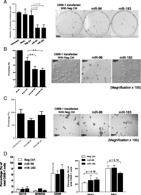
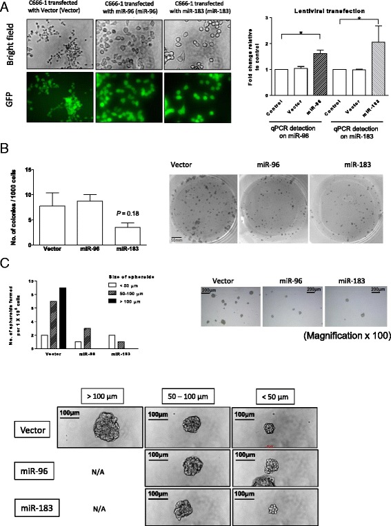
Methods: MiR-96 and miR-183 expression in NPC CSCs was detected by qRT-PCR. Transient and stable transfection was performed in EBV-positive NPC C666-1 cells to examine the effects of ectopic expression of miR-96 and miR-183 on repressing cell growth and CSC properties. Anchorage-dependent (colony formation) and anchorage-independent (tumor sphere formation) growths of these miR-96 and miR-183 expressing cells were determined. Expression of multiple CSC markers and related molecules were accessed by flow cytometry and Western blotting. The tumorigenicity of the stable miR-96- and miR-183-transfected NPC cells was examined in an in vivo nude mice model.

Results: Downregulation of miR-96 and miR-183 was confirmed in NPC spheroids. Using transient or stable transfection, we showed that ectopic expression of miR-96 and miR-183 suppressed cell growth and tumor sphere formation in NPC. Reduced NICD3 and NICD4 in miR-96- and miR-183-expressing NPC cells suggests the involvement of the NOTCH signaling pathway in their tumor suppressive function. Finally, we showed that the tumorigenicity of cells stably expressing miR-183 was significantly inhibited in the in vivo nude mice model.

Conclusions: miR-183 is a tumor-suppressive miRNA in EBV-associated NPC. Its abilities to suppress CSC properties in vitro and effectively reduce tumor growth in vivo shed light on its role as a potential therapeutic target.

Keywords: Cancer stem-like cells; Epstein-Barr virus; NOTCH; Nasopharyngeal carcinoma; microRNA.


Figures

Fig. 1
Fig. 1
Down-regulation of miR-96 and miR-183 in NPC sphere-forming cells. The expression of miR-96, miR-182, and miR-183 in sphere-forming and parental C666-1 cells was determined by qRT-PCR. The histogram shows the fold changes of miRNA expressions between sphere-forming and parental C666-1 cells. Expression of miR-96 and miR-183 was significantly decreased in sphere-forming C666-1 cells. No significant difference in miR-182 expression was found. Student’s t-test was used to determine statistical significance between the two groups (n = 3, **P < 0.01, ***P < 0.001)
Fig. 2
Fig. 2
Transient expression of miR-96 and miR-183 inhibits colony and tumor sphere formation in NPC cells. The effects of transient overexpression of miR-96 and mi-183 on NPC cell growth were examined. a Colony formation assay of C666-1 cells with miR-96 and miR-183 overexpression. The numbers of colonies formed by C666-1 cells transfected with miR-96 and miR-183 were obviously lower than those of the negative control (n = 3, P = 0.08). b Tumor sphere formation in C666-1 cells expressing miR-96 and miR-183 was significantly inhibited when compared to that of the negative control (n = 3, **P < 0.01). Representative photos of tumor spheres are shown in the right panel (magnification × 100). c No change in tumor sphere formation in the C666-1 cells co-transfected with miR-96 or miR-183 and the corresponding anti-miR inhibitors was observed when compared to negative controls (n = 3, all P > 0.05). Representative photos of tumor spheres are shown in the right panel (magnification × 100). d Tumor cells expressing OCT4, NANOG, CD44, SOX2, and BMI1 were quantified by flow cytometry analysis. The histogram shows the percentage of cells expressing these proteins in C666-1 cells transfected with miR-96 and miR-183 and that of the negative control. No significant changes in OCT4, NANOG, CD44, SOX2, and BMI1 expression were observed in the cells transfected with miR-96 and miR-183 (n = 3, all P > 0.05). Student’s t-test was used to determine statistical significance between the two groups
Fig. 3
Fig. 3
Effects of stable overexpression of miR-96 and miR-183 in C666-1. a C666-1 cells stably expressing miR-96 and miR-183 were established by lentiviral transfection. Left panel: Transfection efficiency of the transfected cells expressing green fluorescence protein (GFP) was determined under microscopy. Representative photos show that over 90 % of the cells are visualized in green. Right panel: The expression of miR-96 or miR-186 in the stably transfected cells was confirmed by qRT-PCR. Histograms confirmed elevated expressions of miR-96 and miR-183 in the respective cells stably transfected with miR-96 and miR-183 (n = 3, all *P < 0.05). The effects of miR-96 and miR-183 overexpression in NPC cells were assessed by their (b) colony-forming and (c) sphere-forming capacities. b The histogram shows the number of colonies formed in the C666-1 cells expressing miR-96 or miR-183 compared to that of the vector control. The colony number of C666-1 cells expressing miR-183 is obviously lower than that of the control (n = 3, P = 0.18). However, no aberration was observed in colony formation between cells stably transfected with miR-96 and those with the vector. Representative photos of the colonies are shown in the right panel. c Both the number and size of the tumor spheres were reduced in the C666-1 cells expressing miR-96 and miR-183. The number of tumor spheres in the C666-1 cells stably expressing miR-96 and miR-183 and the vector control is illustrated in the histogram according to the sizes of the tumor spheres formed (<50 μm, 50–100 μm, > 100 μm) (mean data from 6 wells of a 6-well plate per group). C666-1 cells stably expressing miR-96 and miR-183 failed to form tumor spheres of diameter larger than 100 μm. Representative photos of the tumor spheres are shown in the right panel (magnification × 100). Lower panel: Representative photomicrographs reveal the difference in size of the spheres between each treatment group
Fig. 4
Fig. 4
Overexpression of miR-183 inhibits stemness and tumorigenic potential in C666-1 cells. a The expression of stem cell transcription factors NICD3, NICD4, and CYCLIND D1 in the C666-1 cells stably expressing miR-96 and miR-183 was detected by Western blot. Reduced expression of SOX2, OCT4, NICD3, and NICD4 was found in the C666-1 cells with overexpression of miR-96 or miR-183. Similar BMI and CYCLIN D1 expression was observed in the C666-1 cells stably expressing miR-96 and miR-18 and the vector control. Beta-actin was used as the internal loading control. The results were quantified and the histogram shows the fold changes between stable transfected cells and the vector control. b The tumorigenic potential of the C666-1 cells stably expressing miR-96 and miR-183 was assessed by tumor growth in an in vivo nude mice model. Significant inhibition of tumor formation was observed in the C666-1 cells stably expressing miR-183 (**P < 0.01) but not in the cells stably expressing miR-96. Photos reveal the appearances and sizes of the tumors in each treatment group. Student’s t-test was used to determine statistical significance between the two groups (n = 3, **P < 0.01)

References

    1. Cao SM, Simons MJ, Qian CN. The prevalence and prevention of nasopharyngeal carcinoma in China. Chin J Cancer. 2011;30(2):114–119. doi: 10.5732/cjc.010.10377. - DOI - PMC - PubMed
    1. Lo KW, Chung GT, To KF. Deciphering the molecular genetic basis of NPC through molecular, cytogenetic, and epigenetic approaches. Semin Cancer Biol. 2012;22(2):79–86. doi: 10.1016/j.semcancer.2011.12.011. - DOI - PubMed
    1. Lee AW, Lin JC, Ng WT. Current management of nasopharyngeal cancer. Semin Radiat Oncol. 2012;22(3):233–244. doi: 10.1016/j.semradonc.2012.03.008. - DOI - PubMed
    1. Li F, Tiede B, Massague J, Kang Y. Beyond tumorigenesis: cancer stem cells in metastasis. Cell Res. 2007;17(1):3–14. doi: 10.1038/sj.cr.7310118. - DOI - PubMed
    1. Tu SM, Lin SH, Logothetis CJ. Stem-cell origin of metastasis and heterogeneity in solid tumours. Lancet Oncol. 2002;3(8):508–513. doi: 10.1016/S1470-2045(02)00820-3. - DOI - PubMed

Publication types

MeSH terms