

Reliability of measurements on lateral ankle radiographs
- PMID: 27431806
- PMCID: PMC4949875
- DOI: 10.1186/s12891-016-1150-4
Reliability of measurements on lateral ankle radiographs
Abstract
Background: The aims of our study were to evaluate the validation of measurement of weight-bearing lateral radiographs. Two hypotheses were tested: the measurements on the lateral radiographs are reliable, and a theoretical limit could be identified when a surgeon can "eyeball" an incongruous ankle joint on lateral radiographs.
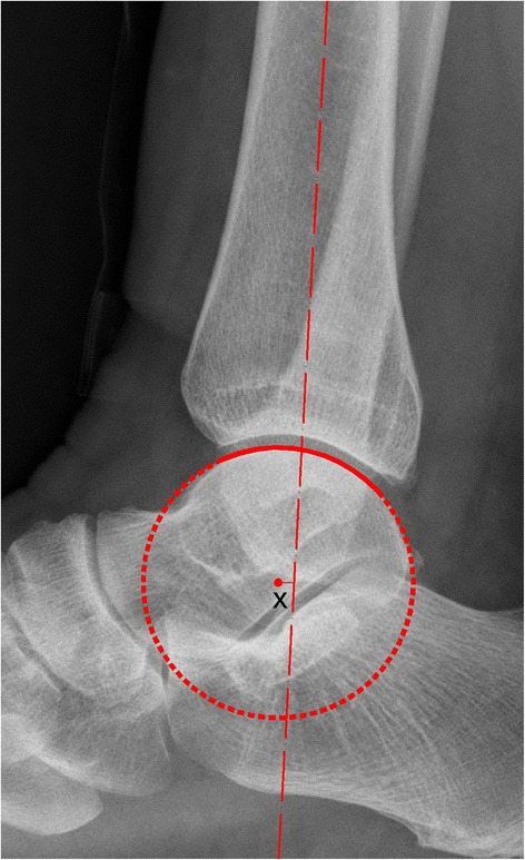
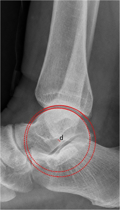
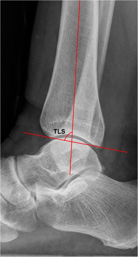
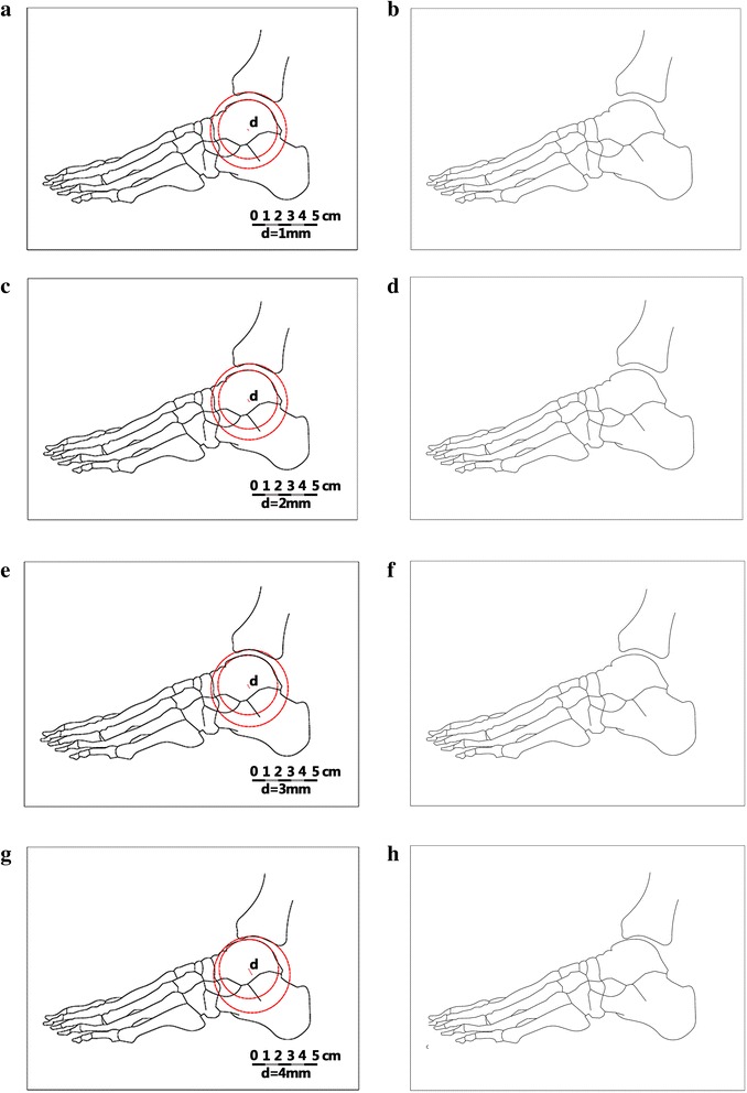
Methods: To test the first hypothesis, 3 experienced ankle surgeons evaluated 50 normal weight-bearing lateral radiographs of patients. The measurements assessed were the tibial lateral surface angle (TLS), the distance from the center of the talar joint circle to the longitudinal axis of the tibia (x) and the displacement from the center of the talar articular joint circle to the center of the distal tibia articular joint circle (d). To test the second hypothesis, we used CAD software to create schematic diagrams on which lateral radiographs of the ankle joint were not parallel (d = 1, 2, 3, 4 mm). Five experienced ankle surgeons were asked to judge whether the ankle articular surfaces were parallel. Intraobserver reliability was determined using the intraclass correlation coefficients (ICCs) and interobserver agreement by the Kendall coefficient of concordance.
Results: First, the intraobserver reliability was high (Cronbach's alpha >0.80) with regard to radiographic measurements according to the ICC. Significant interobserver disagreement was found (Kendall tauB, p < 0.01) using the Kendall concordance coefficient. Second, when the d-value was 4 mm, all the observers identified the incongruous ankle joint at two separate times.
Conclusions: Consultation with experienced foot and ankle surgeons and precise definitions for lateral measurement assessments do not guarantee a high level of agreement. Surgeons can observe an incongruous ankle joint on lateral radiographs when the d-value is 4 mm.
Keywords: Ankle; Interobserver study; Lateral radiographs; Reliability.
Figures
Similar articles
-
Reliability of the Radiographic Sagittal and Frontal Tibiotalar Alignment after Ankle Arthrodesis.PLoS One. 2016 Apr 28;11(4):e0154224. doi: 10.1371/journal.pone.0154224. eCollection 2016. PLoS One. 2016. PMID: 27124403 Free PMC article.
-
Observer reliability in ankle and calcaneocuboid stress radiography.Am J Sports Med. 2008 Jun;36(6):1143-9. doi: 10.1177/0363546507313091. Epub 2008 Feb 20. Am J Sports Med. 2008. PMID: 18287594
-
Lateral talar station: a clinically reproducible measure of sagittal talar position.Foot Ankle Int. 2013 Dec;34(12):1669-76. doi: 10.1177/1071100713500489. Epub 2013 Aug 21. Foot Ankle Int. 2013. PMID: 23966113
-
Understanding Radiographic Measurements Used in Foot and Ankle Surgery.J Am Acad Orthop Surg. 2022 Jan 15;30(2):e139-e154. doi: 10.5435/JAAOS-D-20-00189. J Am Acad Orthop Surg. 2022. PMID: 34768261 Review.
-
Foot and ankle measurements on cone beam weightbearing computed tomography.J Biol Regul Homeost Agents. 2020 May-Jun;34(3 Suppl. 2):23-32. ADVANCES IN MUSCULOSKELETAL DISEASES AND INFECTIONS - SOTIMI 2019. J Biol Regul Homeost Agents. 2020. PMID: 32856436 Review.
Cited by
-
Ankle ultrasound for detecting anterior talofibular ligament tear using operative finding as reference standard: a systematic review and meta-analysis.Eur J Trauma Emerg Surg. 2020 Feb;46(1):73-81. doi: 10.1007/s00068-019-01169-3. Epub 2019 Jun 11. Eur J Trauma Emerg Surg. 2020. PMID: 31187159
-
Torsional Deformity Significantly Impacts Lateral Ankle Radiographic Imaging Parameters.Cureus. 2024 Apr 29;16(4):e59292. doi: 10.7759/cureus.59292. eCollection 2024 Apr. Cureus. 2024. PMID: 38813268 Free PMC article.
-
Does a rupture of the lateral ankle ligament need to be repaired in supination-adduction type II (OTA/AO 44A2) fractures?Arch Orthop Trauma Surg. 2024 Jan;144(1):229-237. doi: 10.1007/s00402-023-05044-0. Epub 2023 Oct 15. Arch Orthop Trauma Surg. 2024. PMID: 37838982
-
Influence of the ankle position and X-ray beam angulation on the projection of the posterior facet of the subtalar joint.Skeletal Radiol. 2019 Oct;48(10):1581-1589. doi: 10.1007/s00256-019-03220-1. Epub 2019 Apr 27. Skeletal Radiol. 2019. PMID: 31030252
References
-
- Weber BG, Simpson LA. Corrective lengthening osteotomy of the fibula. Clin Orthop Relat Res. 1985;199:61–67. - PubMed
Publication types
MeSH terms
LinkOut - more resources
Full Text Sources
Other Literature Sources
Miscellaneous
