

Characterization of fracture behavior of human atherosclerotic fibrous caps using a miniature single edge notched tensile test
- PMID: 27431877
- PMCID: PMC5012960
- DOI: 10.1016/j.actbio.2016.07.027
Characterization of fracture behavior of human atherosclerotic fibrous caps using a miniature single edge notched tensile test
Abstract
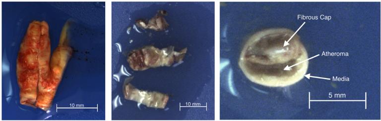
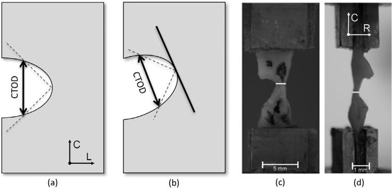
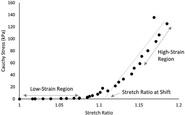
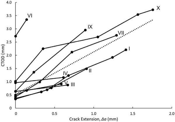
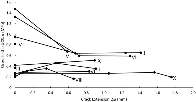
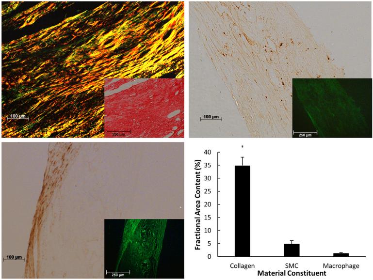
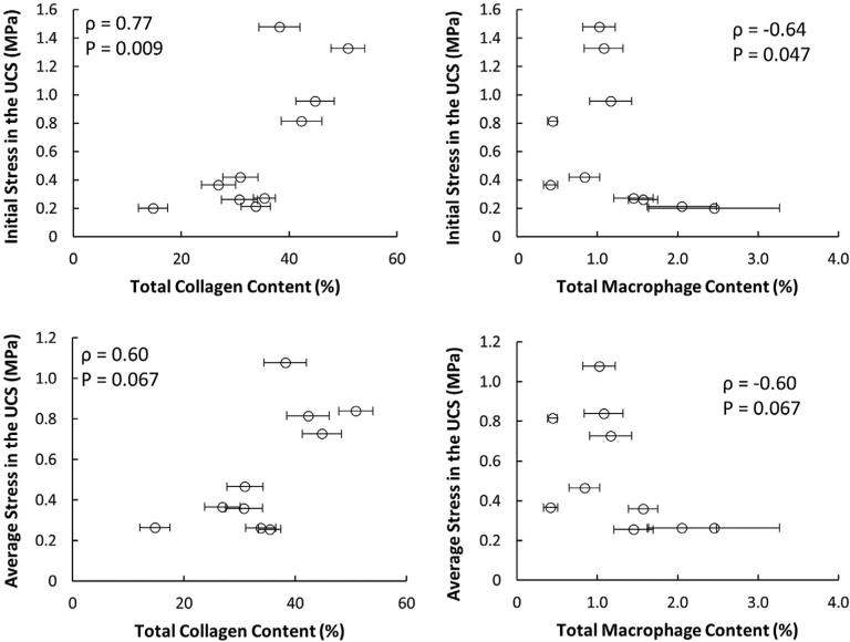
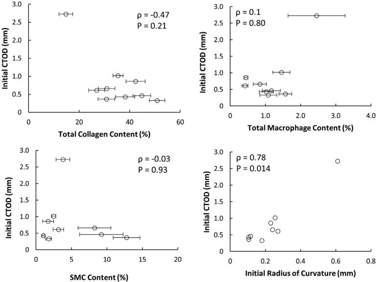
One well-established cause of ischemic stroke is atherosclerotic plaque rupture in the carotid artery. Rupture occurs when a tear in the fibrous cap exposes highly thrombogenic material in the lipid core. Though some fibrous cap material properties have been measured, such as ultimate tensile strength and stress-strain responses, there has been very little, if any, data published regarding the fracture behavior of atherosclerotic fibrous caps. This study aims to characterize the qualitative and quantitative fracture behavior of human atherosclerotic plaque tissue obtained from carotid endarterectomy samples using two different metrics. Uniaxial tensile experiments along with miniature single edge notched tensile (MSENT) experiments were performed on strips of isolated fibrous cap. Crack tip opening displacement (CTOD) and stress in the un-cracked segment (UCS) were measured at failure in fibrous cap MSENT specimens subjected to uniaxial tensile loading. Both CTOD and the degree of crack blunting, measured as the radius of curvature of the crack tip, increased as tearing propagated through the tissue. Higher initial stress in the UCS is significantly correlated with higher collagen content and lower macrophage content in the fibrous cap (ρ=0.77, P=0.009; ρ=-0.64, P=0.047; respectively). Trends in the data show that higher CTOD is inversely related to collagen content, though the sample size in this study is insufficient to statistically substantiate this relationship. To the authors' knowledge, this is the pioneering study examining the fracture behavior of fibrous caps and the first use of the CTOD metric in vascular tissue.
Statement of significance: A tear in the fibrous cap of atherosclerotic plaque can lead to ischemic stroke or myocardial infarction. While there is some information in the literature regarding quantitative measures of fibrous cap failure, there is little information regarding the behavior of the tissue during failure. This study examines the failure behavior of fibrous caps both qualitatively, by examining how and where the tissue fails, and quantitatively, by measuring (a) crack tip opening displacement (CTOD) in vascular tissue for the first time and (b) uniaxial stress in the un-cracked segment (UCS). This study shows that both metrics should be evaluated when assessing plaque vulnerability.
Keywords: Atherosclerosis; Crack blunting; Crack tip opening displacement (CTOD); Fracture mechanics; Plaque rupture.
Copyright © 2016 Acta Materialia Inc. Published by Elsevier Ltd. All rights reserved.
Figures

Similar articles
-
An investigation into the critical role of fibre orientation in the ultimate tensile strength and stiffness of human carotid plaque caps.Acta Biomater. 2021 Apr 1;124:291-300. doi: 10.1016/j.actbio.2021.02.008. Epub 2021 Feb 8. Acta Biomater. 2021. PMID: 33571712
-
A uni-extension study on the ultimate material strength and extreme extensibility of atherosclerotic tissue in human carotid plaques.J Biomech. 2015 Nov 5;48(14):3859-67. doi: 10.1016/j.jbiomech.2015.09.037. Epub 2015 Oct 21. J Biomech. 2015. PMID: 26472304 Free PMC article.
-
Local characterization of collagen architecture and mechanical properties of tissue engineered atherosclerotic plaque cap analogs.Acta Biomater. 2025 Mar 1;194:185-193. doi: 10.1016/j.actbio.2025.01.035. Epub 2025 Jan 22. Acta Biomater. 2025. PMID: 39855375
-
Contemporary carotid imaging: from degree of stenosis to plaque vulnerability.J Neurosurg. 2016 Jan;124(1):27-42. doi: 10.3171/2015.1.JNS142452. Epub 2015 Jul 31. J Neurosurg. 2016. PMID: 26230478 Review.
-
Uniaxial tensile testing approaches for characterisation of atherosclerotic plaques.J Biomech. 2014 Mar 3;47(4):793-804. doi: 10.1016/j.jbiomech.2014.01.017. Epub 2014 Jan 14. J Biomech. 2014. PMID: 24508324 Review.
Cited by
-
Diffusion tensor imaging and arterial tissue: establishing the influence of arterial tissue microstructure on fractional anisotropy, mean diffusivity and tractography.Sci Rep. 2020 Nov 26;10(1):20718. doi: 10.1038/s41598-020-77675-x. Sci Rep. 2020. PMID: 33244026 Free PMC article.
-
LIFU-responsive nanomedicine enables acoustic droplet vaporization-induced apoptosis of macrophages for stabilizing vulnerable atherosclerotic plaques.Bioact Mater. 2022 Mar 3;16:120-133. doi: 10.1016/j.bioactmat.2022.02.022. eCollection 2022 Oct. Bioact Mater. 2022. PMID: 35386311 Free PMC article.
-
Fracture properties of porcine versus human thoracic aortas from tricuspid/bicuspid aortic valve patients via symmetry-constraint Compact Tension testing.Sci Rep. 2025 Jan 3;15(1):667. doi: 10.1038/s41598-024-83233-6. Sci Rep. 2025. PMID: 39753641 Free PMC article.
-
Preliminary Investigation of the Mechanical Anisotropy of the Normal Human Corneal Stroma.J Ophthalmol. 2018 Oct 17;2018:5392041. doi: 10.1155/2018/5392041. eCollection 2018. J Ophthalmol. 2018. PMID: 30416826 Free PMC article.
-
[Numerical simulation study of fracture mechanics of the atherosclerotic plaque].Sheng Wu Yi Xue Gong Cheng Xue Za Zhi. 2021 Dec 25;38(6):1097-1102. doi: 10.7507/1001-5515.202106077. Sheng Wu Yi Xue Gong Cheng Xue Za Zhi. 2021. PMID: 34970892 Free PMC article. Chinese.
References
-
- Heron M. Deaths: Leading causes for 2010. Nat. Vital Stat. Rep. 2013;62 - PubMed
-
- Go AS, Mozaffarian D, Roger VL, Benjamin EJ, Berry JD, Blaha MJ, Dai S, Ford ES, Fox CS, Franco S, Fullerton HJ, Gillespie C, Hailpern SM, Heit JA, Howard VJ, Huffman MD, Judd SE, Kissela BM, Kittner SJ, Lackland DT, Lichtman JH, Lisabeth LD, Mackey RH, Magid DJ, Marcus GM, Marelli A, Matchar DB, McGuire DK, Mohler ER, III, Moy CS, Mussolino ME, Neumar RW, Nichol G, Pandey DK, Paynter NP, Reeves MJ, Sorlie PD, Stein J, Towfighi A, Turan TN, Virani SS, Wong ND, Woo D, Turner MB. Heart disease and stroke statistics–2014 update: a report from the American Heart Association. Circulation. 2014;128 http://dx.doi.org/10.1161/01.cir.0000441139.02102.80. - DOI - PMC - PubMed
-
- Petty GW, Brown RD, Whisnant JP, Sicks JD, O'Fallon WM, Wiebers DO. Ischemic stroke subtypes: a population-based study of incidence and risk factors. Stroke. 1999;30:2513–2516. http://dx.doi.org/10.1161/01.STR.30.12.2513. - DOI - PubMed
-
- Burke AP, Kolodgie FD, Farb A, Weber DK, Malcom GT, Smialek J, Virmani R. Healed plaque ruptures and sudden coronary death: Evidence that subclinical rupture has a role in plaque progression. Circulation. 2001;103:934–940. http://dx.doi.org/10.1161/01.CIR.103.7.934. - DOI - PubMed
-
- Bentzon JF, Otsuka F, Virmani R, Falk E. Mechanisms of plaque formation and rupture. Circ. Res. 2014;114:1852–1866. http://dx.doi.org/10.1161/CIRCRESAHA.114.302721. - DOI - PubMed
Publication types
MeSH terms
Grants and funding
LinkOut - more resources
Full Text Sources
Other Literature Sources
Research Materials
Miscellaneous
