Coronal tibial slope is associated with accelerated knee osteoarthritis: data from the Osteoarthritis Initiative
- PMID: 27432004
- PMCID: PMC4950083
- DOI: 10.1186/s12891-016-1158-9
Coronal tibial slope is associated with accelerated knee osteoarthritis: data from the Osteoarthritis Initiative
Abstract
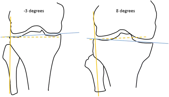
Background: Accelerated knee osteoarthritis may be a unique subset of knee osteoarthritis, which is associated with greater knee pain and disability. Identifying risk factors for accelerated knee osteoarthritis is vital to recognizing people who will develop accelerated knee osteoarthritis and initiating early interventions. The geometry of an articular surface (e.g., coronal tibial slope), which is a determinant of altered joint biomechanics, may be an important risk factor for incident accelerated knee osteoarthritis. We aimed to determine if baseline coronal tibial slope is associated with incident accelerated knee osteoarthritis or common knee osteoarthritis.
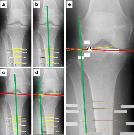
Methods: We conducted a case-control study using data and images from baseline and the first 4 years of follow-up in the Osteoarthritis Initiative. We included three groups: 1) individuals with incident accelerated knee osteoarthritis, 2) individuals with common knee osteoarthritis progression, and 3) a control group with no knee osteoarthritis at any time. We did 1:1:1 matching for the 3 groups based on sex. Weight-bearing, fixed flexion posterior-anterior knee radiographs were obtained at each visit. One reader manually measured baseline coronal tibial slope on the radiographs. Baseline femorotibial angle was measured on the radiographs using a semi-automated program. To assess the relationship between slope (predictor) and incident accelerated knee osteoarthritis or common knee osteoarthritis (outcomes) compared with no knee osteoarthritis (reference outcome), we performed multinomial logistic regression analyses adjusted for sex.
Results: The mean baseline slope for incident accelerated knee osteoarthritis, common knee osteoarthritis, and no knee osteoarthritis were 3.1(2.0), 2.7(2.1), and 2.6(1.9); respectively. A greater slope was associated with an increased risk of incident accelerated knee osteoarthritis (OR = 1.15 per degree, 95 % CI = 1.01 to 1.32) but not common knee osteoarthritis (OR = 1.04, 95 % CI = 0.91 to 1.19). These findings were similar when adjusted for recent injury. Among knees with varus malalignment a greater slope increases the odds of incident accelerated knee osteoarthritis; there is no significant relationship between slope and incident accelerated knee osteoarthritis among knees with normal alignment.
Conclusions: Coronal tibial slope, particularly among knees with malalignment, may be an important risk factor for incident accelerated knee osteoarthritis.
Keywords: Alignment; Bone; Knee; Osteoarthritis; Radiography.
Figures


References
-
- Neogi T, Niu J, Duryea J, Lynch J, Zhang Y. Identifying trajectories of medial joint-space width loss and associated risk factors. Osteoarthritis Cartilage. 2012;20(Supplement 1):S182–3. doi: 10.1016/j.joca.2012.02.281. - DOI
-
- Driban JB, Price LL, Eaton CB, Lu B, Lo GH, Lapane KL, McAlindon TE. Individuals with incident accelerated knee osteoarthritis have greater pain than those with common knee osteoarthritis progression: data from the Osteoarthritis Initiative. Clin Rheumatol. 2015;35:1565–71. doi: 10.1007/s10067-015-3128-2. - DOI - PMC - PubMed
Publication types
MeSH terms
Grants and funding
LinkOut - more resources
Full Text Sources
Other Literature Sources

