. 2016 Aug;32(4):349-62.
doi: 10.1007/s12264-016-0047-4. Epub 2016 Jul 18.

Echinacoside Protects Against MPP(+)-Induced Neuronal Apoptosis via ROS/ATF3/CHOP Pathway Regulation

Affiliations

Echinacoside Protects Against MPP(+)-Induced Neuronal Apoptosis via ROS/ATF3/CHOP Pathway Regulation

Qing Zhao et al. Neurosci Bull. 2016 Aug.

Abstract

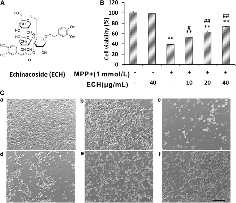
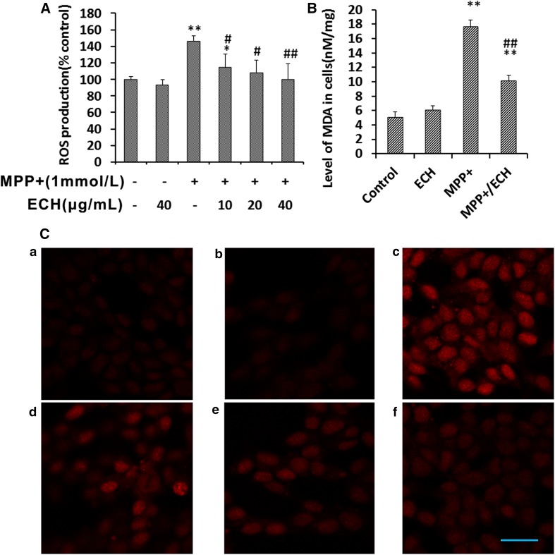
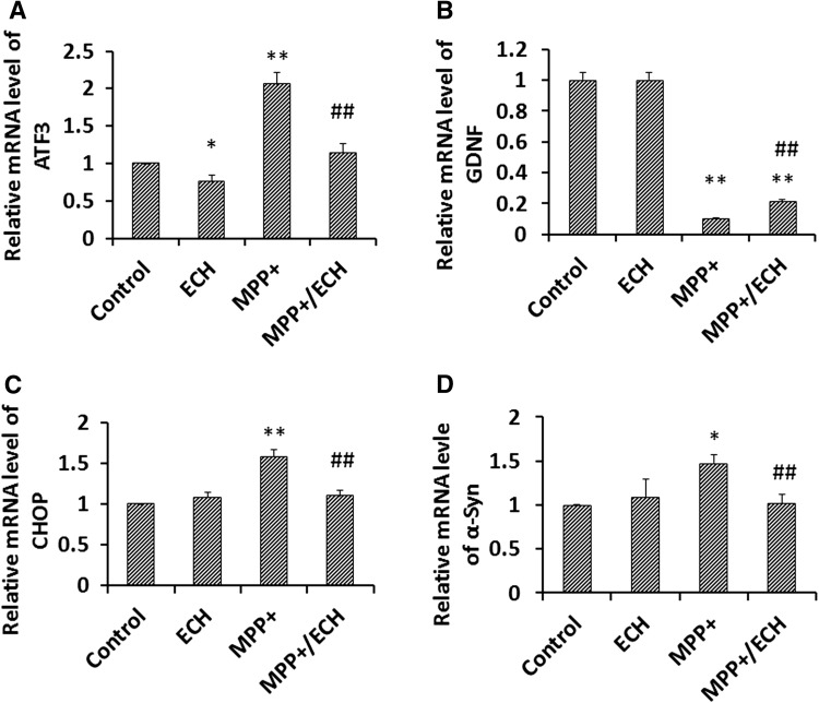
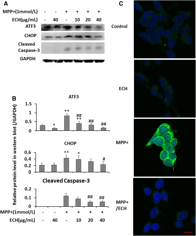
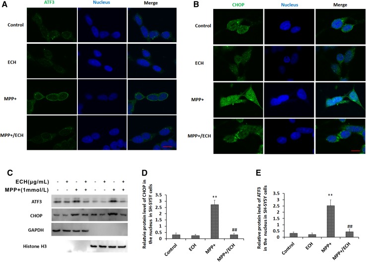
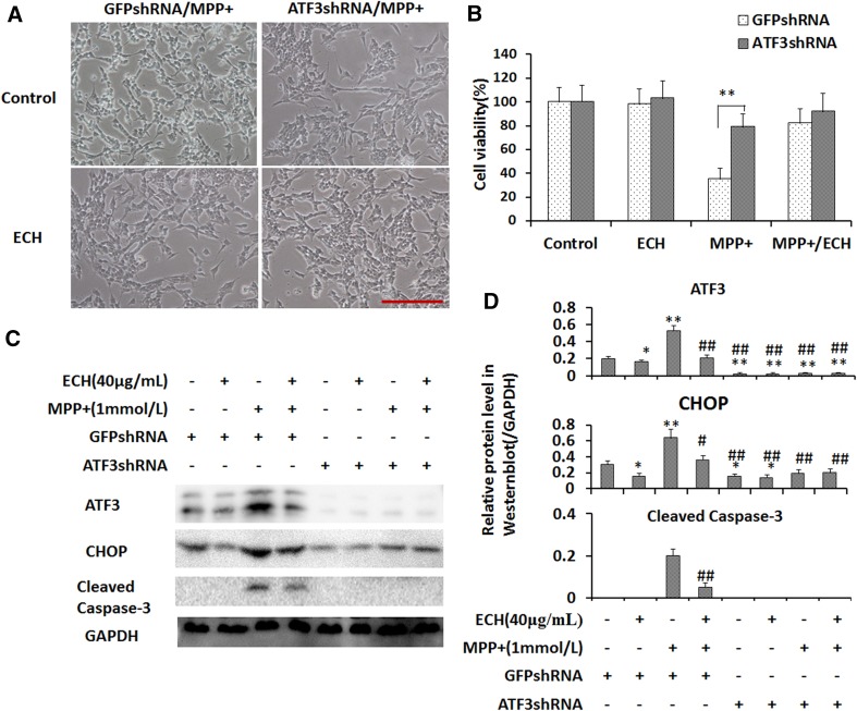
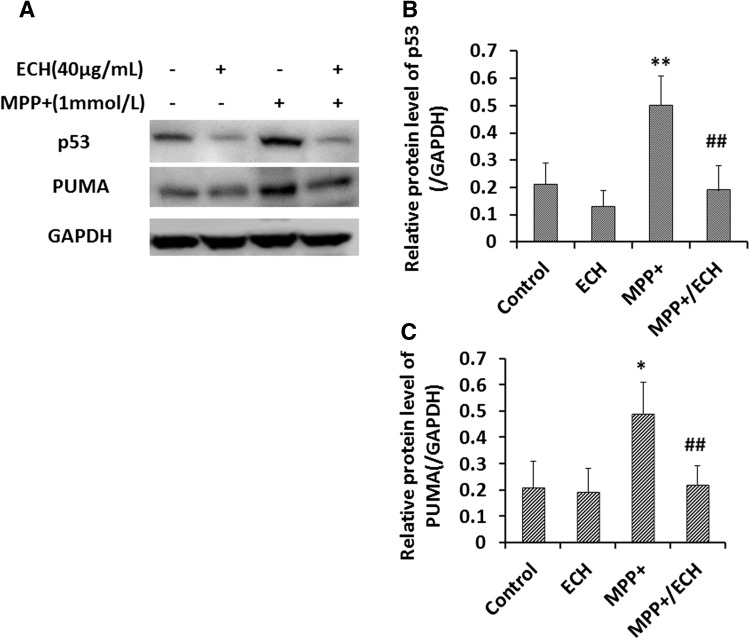
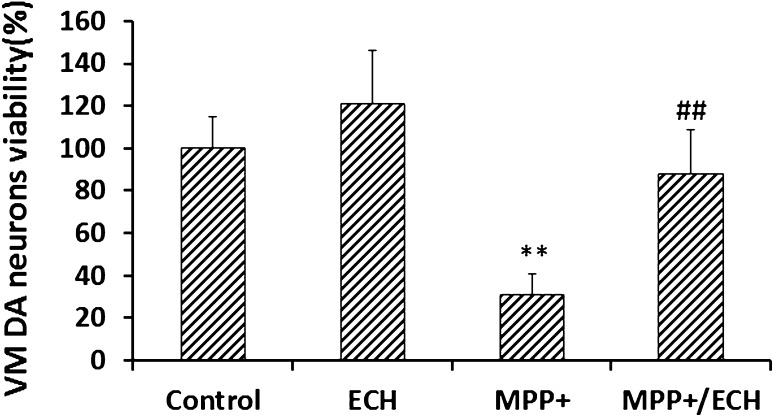
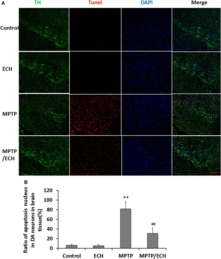
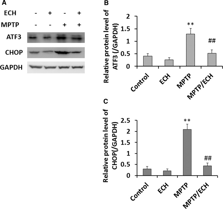
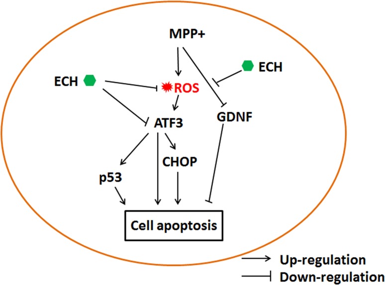
Echinacoside (ECH) is protective in a mouse model of Parkinson's disease (PD) induced by 1-methyl-4-phenylpyridinium ion (MPP(+)). To investigate the mechanisms involved, SH-SY5Y neuroblastoma cells were treated with MPP(+) or a combination of MPP(+) and ECH, and the expression of ATF3 (activating transcription factor 3), CHOP (C/EBP-homologous protein), SCNA (synuclein alpha), and GDNF (glial cell line-derived neurotrophic factor) was assessed. The results showed that ECH significantly improved cell survival by inhibiting the generation of MPP(+)-induced reactive oxygen species (ROS). In addition, ECH suppressed the ROS and MPP(+)-induced expression of apoptotic genes (ATF3, CHOP, and SCNA). ECH markedly decreased the MPP(+)-induced caspase-3 activity in a dose-dependent manner. ATF3-knockdown also decreased the CHOP and cleaved caspase-3 levels and inhibited the apoptosis induced by MPP(+). Interestingly, ECH partially restored the GDNF expression that was down-regulated by MPP(+). ECH also improved dopaminergic neuron survival during MPP(+) treatment and protected these neurons against the apoptosis induced by MPTP. Taken together, these data suggest that the ROS/ATF3/CHOP pathway plays a critical role in mechanisms by which ECH protects against MPP(+)-induced apoptosis in PD.

Keywords: 1-Methyl-4-phenylpyridinium ion; ATF3; CHOP; Echinacoside; Parkinson’s disease; Reactive oxygen species.


Figures

Fig. 1
Fig. 1
Echinacoside inhibits MPP+-induced apoptosis in SH-SY5Y cells. A Chemical structure of echinacoside (Pusi Biotechnology Company). B Effects of echinacoside on viability with or without MPP+. SH-SY5Y cells were plated into 96-well plates, incubated overnight, and then fed fresh medium each day with the indicated drugs; viability was analyzed after 3 days. Data from three independent experiments were normalized to the results from control cells and are presented as mean ± SD (n = 6; **P < 0.01 vs control, # P < 0.05, ## P < 0.01 vs MPP+ treatment). C Light microscopic images of morphological changes induced by MPP+ or MPP+ and ECH. a, PBS control; b, 40 μg/mL ECH; c, 1 mmol/L MPP+; d, 1 mmol/L MPP+ and 10 μg/mL ECH; e, 1 mmol/L MPP+ and 20 μg/mL ECH; f, 1 mmol/L MPP+ and 40 μg/mL ECH (scale bar, 200 μm).
Fig. 2
Fig. 2
Echinacoside decreases MPP+-induced ROS production in SH-SY5Y cells. A Effects of ECH on MPP+-induced ROS products. Cells were seeded into 96-well plates, cultured overnight, and then treated as indicated for 24 h. DCFH-DA (10 μmol/L) was used to analyze ROS levels. Data were normalized to the control group and are presented as mean ± SD (n = 6; *P < 0.05, **P < 0.01; # P < 0.05, ## P < 0.01 vs MPP+ treatment). B ECH blocked the MPP+-induced increase of MDA expression. Cells were incubated with indicated drugs for 24 h. Control, PBS; ECH, 40 μg/mL; MPP+, 1 mmol/L; MPP+/ECH, 1 mmol/L MPP+ and 40 μg/mL ECH. Data were normalized to the control group and are presented as mean ± SD (n = 6; **P < 0.01 vs control; # P < 0.05, ## P < 0.01 vs MPP+ treatment). C Representative fluorescence microscopic images of ROS expression in cells treated with different drugs for 24 h, then incubated with 10 μmol/L DCFH-DA for 20 min. a, PBS control; b, 40 μg/mL ECH; c, 1 mmol/L MPP+; d, 1 mmol/L MPP+ and 10 μg/mL ECH; e, 1 mmol/L MPP+ and 20 μg/mL ECH; f, 1 mmol/L MPP+ and 40 μg/mL ECH (scale bar, 25 μm).
Fig. 3
Fig. 3
Echinacoside regulates expression of ROS stress- and survival-related genes induced by MPP+ in SH-SY5Y cells. qRT-PCR analysis of target mRNA expression (A ATF3; B GDNF; C CHOP; D α-Syn). Cells plated in 6-well plates and cultured for 24 h were then treated as indicated for 24 h. Data from three independent experiments were normalized to the PBS control group. Data are presented as mean ± SD (n = 3; Control, PBS; ECH, 40 μg/mL; MPP+, 1 mmol/L; MPP+/ECH, 1 mmol/L MPP+ and 40 μg/mL ECH; *P < 0.05, **P < 0.01 vs control; # P < 0.05, ## P < 0.01 vs ECH).
Fig. 4
Fig. 4
Echinacoside downregulates ROS stress-related and pro-apoptotic proteins in SH-SY5Y cells. A Western blots of ATF3, CHOP, and cleaved caspase-3 protein from cells treated as indicated for 24 h. B Statistics of relative protein levels as in (A). Data are presented as mean ± SD of the ratio of the indicated protein to GAPDH (n = 3). C Immunofluorescence images showing that ECH suppressed the caspase-3 activation induced by MPP+ (Control, PBS; ECH, 40 μg/mL; MPP+, 1 mmol/L; MPP+/ECH, 1 mmol/L MPP+ and 40 μg/mL ECH; scale bar, 10 μm).
Fig. 5
Fig. 5
Echinacoside decreases nuclear expression of ATF3 and CHOP proteins induced by MPP+ in SH-SY5Y cells. A, B Images of cells stained for endogenous ATF3 (green in A) and CHOP (green in B). Alexa®488-goat anti-rabbit IgG and Hoechst 33342 were sequentially used to stain each target protein and nuclei (blue). Scale bar, 10 μm. C Western blots of ATF3 and CHOP proteins in the cytoplasmic and nuclear fractions. D, E Statistics for experiments as in (C). Data are presented as mean ± SD of the ratio of the indicated protein to histone3 (n = 3; **P < 0.01 vs control group, ## P < 0.01 vs MPP+ group).
Fig. 6
Fig. 6
ATF3 plays a critical role in the action of ECH against MPP+-induced apoptosis via the ROS stress pathway in SH-SY5Y cells. A Downregulation of ATF3 attenuates morphological changes induced by MPP+. Light microscopic images of ATF3 knock-down and control cells after 24-h treatment as indicated (Control, PBS and 1 mmol/L MPP+; ECH, 40 μg/mL ECH and 1 mmol/L MPP+, scale bar, 200 μm). B Cell viability normalized to the control group. Data are presented as mean ± SD (n = 6; **P < 0.01). C Western blots of ATF3, CHOP, and cleaved caspase-3 proteins with GAPDH serving as the loading control. D Statistics for experiments as in (C). Data are presented as mean ± SD of the ratio of the indicated protein to GAPDH (n = 3).
Fig. 7
Fig. 7
Echinacoside downregulates p53 and PUMA in SH-SY5Y cells. A Western blots of p53 and PUMA proteins in cells treated as indicated for 24 h. GAPDH served as loading control. B, C Statistics for experiments as in (A). Data are presented as mean ± SD of the ratio of the indicated protein to GAPDH (n = 3; Control, PBS; ECH, 40 μg/mL; MPP+, 1 mmol/L; MPP+/ECH, 1 mmol/L MPP+ and 40 μg/mL ECH).
Fig. 8
Fig. 8
ECH protects primary DA neurons in vitro. Viability of rat primary VM DA neurons treated as indicated. Data from three independent experiments normalized to the results from control cells and presented as mean ± SD (n = 6; **P < 0.01).
Fig. 9
Fig. 9
ECH blocks DA neuronal apoptosis in mouse PD model. A Immunofluorescence and TUNEL assay images of apoptotic TH-positive (TH+) cells in sections through the substantia nigra pars compacta. Nuclei were stained with DAPI (blue). TH+ cells (green) and TH+ and TUNEL-positive nuclei (red) were counted (scale bar, 200 μm). B Ratios of apoptotic DA neurons. Data are presented as mean ± SD (n = 5; **P < 0.01 vs control group; ## P < 0.01 vs MPP+ group).
Fig. 10
Fig. 10
ECH suppresses ATF3 and CHOP expression in mouse PD model. A Western blots of ATF3 and CHOP proteins in lysates of mouse SNc tissue, with GAPDH serving as the loading control. B, C Statistics for experiments as in (A). Data are presented as mean ± SD of the ratio of the indicated protein to GAPDH (n = 5).
Fig. 11
Fig. 11
Proposed mechanisms underlying the protective actions of ECH against MPP+-induced apoptosis in SH-SY5Y cells. ECH attenuates the ROS formation induced by MPP+, and in turn downregulates the expression of ATF3 and CHOP. ECH also directly inhibits ATF3 expression. Another branch shows that ECH promotes the expression of the anti-apoptosis protein, GDNF, in MPP+-treated cells. ATF3 regulates expression of the pro-apoptosis proteins p53 in the PD cell model as well. All of these actions contribute to the protective effect of ECH against apoptosis induced by MPP+.

Similar articles

Cited by

References

    1. Kalia LV, Lang AE. Parkinson’s disease. Lancet. 2015;386:896–902. doi: 10.1016/S0140-6736(14)61393-3. - DOI - PubMed
    1. Lees AJ, Hardy J, Revesz T. Parkinson’s disease. Lancet. 2009;373:2055–2066. doi: 10.1016/S0140-6736(09)60492-X. - DOI - PubMed
    1. Pagonabarraga J, Kulisevsky J, Strafella AP, Krack P. Apathy in Parkinson’s disease: clinical features, neural substrates, diagnosis, and treatment. Lancet Neurol. 2015;14:518–531. doi: 10.1016/S1474-4422(15)00019-8. - DOI - PubMed
    1. Lehri-Boufala S, Ouidja MO, Barbier-Chassefiere V, Henault E, Raisman-Vozari R, Garrigue-Antar L, et al. New roles of glycosaminoglycans in alpha-synuclein aggregation in a cellular model of Parkinson disease. PLoS One. 2015;10:e0116641. doi: 10.1371/journal.pone.0116641. - DOI - PMC - PubMed
    1. Duka T, Duka V, Joyce JN, Sidhu A. Alpha-Synuclein contributes to GSK-3beta-catalyzed Tau phosphorylation in Parkinson’s disease models. FASEB J. 2009;23:2820–2830. doi: 10.1096/fj.08-120410. - DOI - PMC - PubMed

MeSH terms

LinkOut - more resources