

Mass Spectrometric Quantification of N-Linked Glycans by Reference to Exogenous Standards
- PMID: 27432553
- PMCID: PMC5501980
- DOI: 10.1021/acs.jproteome.6b00132
Mass Spectrometric Quantification of N-Linked Glycans by Reference to Exogenous Standards
Abstract
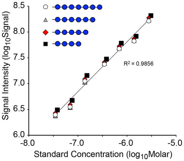
Environmental and metabolic processes shape the profile of glycoprotein glycans expressed by cells, whether in culture, developing tissues, or mature organisms. Quantitative characterization of glycomic changes associated with these conditions has been achieved historically by reductive coupling of oligosaccharides to various fluorophores following release from glycoprotein and subsequent HPLC or capillary electrophoretic separation. Such labeling-based approaches provide a robust means of quantifying glycan amount based on fluorescence yield. Mass spectrometry, on the other hand, has generally been limited to relative quantification in which the contribution of the signal intensity for an individual glycan is expressed as a percent of the signal intensity summed over the total profile. Relative quantification has been valuable for highlighting changes in glycan expression between samples; sensitivity is high, and structural information can be derived by fragmentation. We have investigated whether MS-based glycomics is amenable to absolute quantification by referencing signal intensities to well-characterized oligosaccharide standards. We report the qualification of a set of N-linked oligosaccharide standards by NMR, HPLC, and MS. We also demonstrate the dynamic range, sensitivity, and recovery from complex biological matrices for these standards in their permethylated form. Our results indicate that absolute quantification for MS-based glycomic analysis is reproducible and robust utilizing currently available glycan standards.
Keywords: N-glycan; mass spectrometry; permethylation; quantification; standard.
Conflict of interest statement
The authors declare no competing financial interest.
Figures


Similar articles
-
High-throughput quantitative analysis of total N-glycans by matrix-assisted laser desorption/ionization time-of-flight mass spectrometry.Anal Chem. 2012 Apr 3;84(7):3453-60. doi: 10.1021/ac203440c. Epub 2012 Mar 23. Anal Chem. 2012. PMID: 22455307
-
Providing Bionic Glycome as internal standards by glycan reducing and isotope labeling for reliable and simple quantitation of N-glycome based on MALDI- MS.Anal Chim Acta. 2019 Nov 12;1081:112-119. doi: 10.1016/j.aca.2019.07.003. Epub 2019 Jul 3. Anal Chim Acta. 2019. PMID: 31446948
-
A relative and absolute quantification of neutral N-linked oligosaccharides using modification with carboxymethyl trimethylammonium hydrazide and matrix-assisted laser desorption/ionization time-of-flight mass spectrometry.Anal Biochem. 2008 Aug 1;379(1):45-59. doi: 10.1016/j.ab.2008.04.039. Epub 2008 Apr 27. Anal Biochem. 2008. PMID: 18498755
-
Recent Advances in Labeling-Based Quantitative Glycomics: From High-Throughput Quantification to Structural Elucidation.Proteomics. 2025 Jan;25(1-2):e202400057. doi: 10.1002/pmic.202400057. Epub 2024 Nov 24. Proteomics. 2025. PMID: 39580675 Free PMC article. Review.
-
Two-dimensional HPLC separation with reverse-phase-nano-LC-MS/MS for the characterization of glycan pools after labeling with 2-aminobenzamide.Methods Mol Biol. 2009;534:79-91. doi: 10.1007/978-1-59745-022-5_6. Methods Mol Biol. 2009. PMID: 19277534 Review.
Cited by
-
Glucuronylated core 1 glycans are required for precise localization of neuromuscular junctions and normal formation of basement membranes on Drosophila muscles.Dev Biol. 2018 Apr 15;436(2):108-124. doi: 10.1016/j.ydbio.2018.02.017. Epub 2018 Feb 27. Dev Biol. 2018. PMID: 29499182 Free PMC article.
-
A mutation in SLC37A4 causes a dominantly inherited congenital disorder of glycosylation characterized by liver dysfunction.Am J Hum Genet. 2021 Jun 3;108(6):1040-1052. doi: 10.1016/j.ajhg.2021.04.013. Epub 2021 May 7. Am J Hum Genet. 2021. PMID: 33964207 Free PMC article.
-
COG7 deficiency in Drosophila generates multifaceted developmental, behavioral and protein glycosylation phenotypes.J Cell Sci. 2017 Nov 1;130(21):3637-3649. doi: 10.1242/jcs.209049. Epub 2017 Sep 7. J Cell Sci. 2017. PMID: 28883096 Free PMC article.
-
Reliable N-Glycan Analysis-Removal of Frequently Occurring Oligosaccharide Impurities by Enzymatic Degradation.Molecules. 2023 Feb 15;28(4):1843. doi: 10.3390/molecules28041843. Molecules. 2023. PMID: 36838829 Free PMC article.
-
Neuropathic pain in a Fabry disease rat model.JCI Insight. 2018 Mar 22;3(6):e99171. doi: 10.1172/jci.insight.99171. JCI Insight. 2018. PMID: 29563343 Free PMC article.
References
-
- Geyer H, Geyer R. Strategies for analysis of glycoprotein glycosylation. Biochim Biophys Acta, Proteins Proteomics. 2006;1764(12):1853–69. - PubMed
-
- Ito H, Kaji H, Togayachi A, Azadi P, Ishihara M, Geyer R, Galuska C, Geyer H, Kakehi K, Kinoshita M, Karlsson NG, Jin C, Kato K, Yagi H, Kondo S, Kawasaki N, Hashii N, Kolarich D, Stavenhagen K, Packer NH, Thaysen-Andersen M, Nakano M, Taniguchi N, Kurimoto A, Wada Y, Tajiri M, Yang P, Cao W, Li H, Rudd PM, Narimatsu H. Comparison of analytical methods for profiling N- and O-linked glycans from cultured cell lines: HUPO Human Disease Glycomics/Proteome Initiative multi-institutional study. Glycoconjugate J. 2016;33:405. - PMC - PubMed
-
- Bigge JC, Patel TP, Bruce JA, Goulding PN, Charles SM, Parekh RB. Nonselective and efficient fluorescent labeling of glycans using 2-amino benzamide and anthranilic acid. Anal Biochem. 1995;230(2):229–38. - PubMed
-
- Guile GR, Rudd PM, Wing DR, Prime SB, Dwek RA. A rapid high-resolution high-performance liquid chromatographic method for separating glycan mixtures and analyzing oligosaccharide profiles. Anal Biochem. 1996;240(2):210–26. - PubMed
Publication types
MeSH terms
Substances
Grants and funding
LinkOut - more resources
Full Text Sources
Other Literature Sources
