

Case Reports
doi: 10.1136/bcr-2016-216369.
Intramesosigmoid hernia complicated by strangulated small-bowel obstruction
Affiliations
- PMID: 27432828
- PMCID: PMC4956957
- DOI: 10.1136/bcr-2016-216369
Item in Clipboard
Case Reports
Intramesosigmoid hernia complicated by strangulated small-bowel obstruction
BMJ Case Rep.
.
Abstract
An intramesosigmoid hernia is 1 of the 3 rare types of sigmoid-related hernias that could be complicated by intestinal obstruction. Our patient presented with a clinical picture of intestinal obstruction. CT scan showed features of strangulated small-bowel obstruction secondary to a sigmoid-related hernia. This was confirmed intraoperatively to be an intramesosigmoid hernia. We share the radiological findings with intraoperative surgical correlation and discuss the imaging features described in the literature.
2016 BMJ Publishing Group Ltd.
Figures
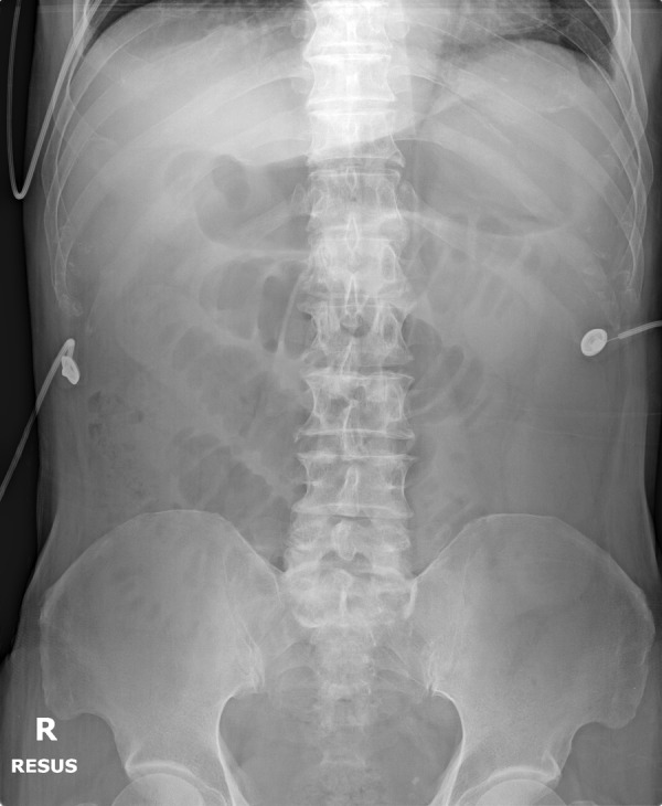
The supine abdominal radiograph of the patient showing several dilated small intestinal loops, suspicious for small-bowel obstruction with a distal transition zone. There is no evidence of overt pneumoperitoneum.
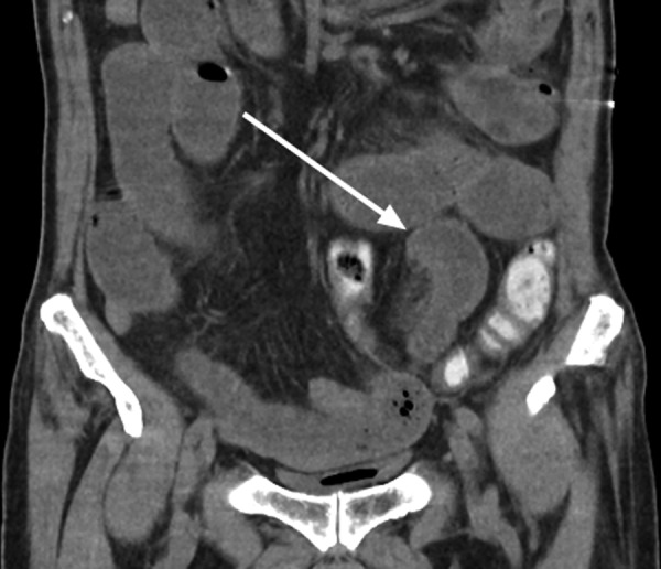
The unenhanced coronal CT image showing a C-shaped appearance of the dilated small bowel (white arrow) with mesenteric engorgement and fat stranding suggestive of strangulation.
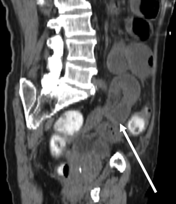
The oblique reformatted CT image better demonstrating the herniated loop of small bowel (white arrow) with a transition zone.
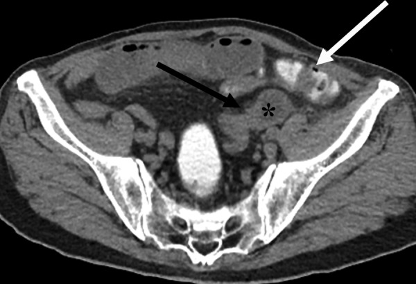
The axial CT image showing the dilated small bowel (*) in an abnormal position, posterolateral to the sigmoid colon that contains intraluminal contrast (white arrow). Its transition zone points medially (black arrow) with a beak-like appearance.
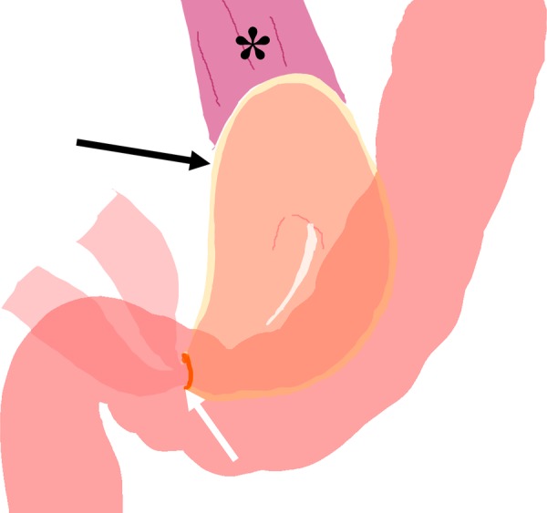
A diagrammatic illustration showing the anatomical relationship of the bowel loops in an intramesosigmoid hernia. The bowel herniates through a defect in the medial leaf of the mesocolon (white arrow) into a sac formed by the lateral leaf of the mesocolon (black arrow). The herniated bowel is found anterior to the left psoas muscle (*) and posterior to the sigmoid.
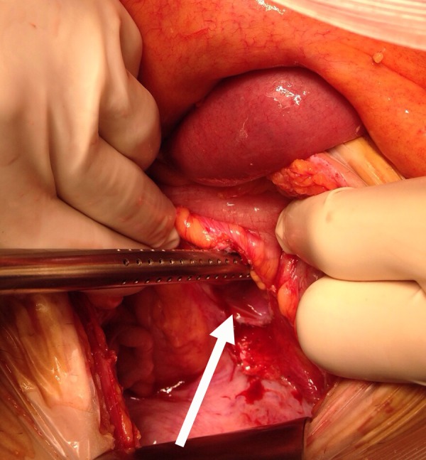
An intraoperative photograph showing the defect in the medial leaf of the mesocolon (white arrow), through which the discoloured ischaemic small bowel had herniated through.
Similar articles
-
Laparoscopic treatment of small bowel strangulation caused by an intramesosigmoid hernia and review of literature.BMJ Case Rep. 2020 Apr 29;13(4):e233627. doi: 10.1136/bcr-2019-233627. BMJ Case Rep. 2020. PMID: 32354762 Free PMC article. Review.
-
Small bowel obstruction due to an intramesosigmoid hernia diagnosed by multidetector row computed tomography: a case report.Osaka City Med J. 2010 Dec;56(2):37-45. Osaka City Med J. 2010. PMID: 21466128
-
Left paraduodenal hernia causing small bowel obstruction in an adolescent patient.J Pediatr Surg. 2009 Dec;44(12):2417-9. doi: 10.1016/j.jpedsurg.2009.09.020. J Pediatr Surg. 2009. PMID: 20006041
-
Education and imaging. Gastrointestinal: a rare cause of small bowel obstruction, paracecal hernia.J Gastroenterol Hepatol. 2015 Mar;30(3):437. doi: 10.1111/jgh.12817. J Gastroenterol Hepatol. 2015. PMID: 25707789 No abstract available.
-
Left paraduodenal hernia causing small bowel obstruction.J Gastrointest Surg. 2014 Jul;18(7):1377-8. doi: 10.1007/s11605-014-2517-1. Epub 2014 Apr 26. J Gastrointest Surg. 2014. PMID: 24771461 Review.
Cited by
-
Intersigmoid Hernia: A Forgotten Diagnosis-A Systematic Review of the Literature over Anatomical, Diagnostic, Surgical, and Medicolegal Aspects.Emerg Med Int. 2020 Jun 1;2020:4891796. doi: 10.1155/2020/4891796. eCollection 2020. Emerg Med Int. 2020. PMID: 32566302 Free PMC article. Review.
-
Laparoscopic treatment of small bowel strangulation caused by an intramesosigmoid hernia and review of literature.BMJ Case Rep. 2020 Apr 29;13(4):e233627. doi: 10.1136/bcr-2019-233627. BMJ Case Rep. 2020. PMID: 32354762 Free PMC article. Review.
-
Small bowel obstruction secondary to intramesosigmoid hernia.BMJ Case Rep. 2019 May 23;12(5):e228766. doi: 10.1136/bcr-2018-228766. BMJ Case Rep. 2019. PMID: 31126929 Free PMC article.
References
Publication types
MeSH terms
LinkOut - more resources
Full Text Sources
Other Literature Sources
Medical