

Mapping the Effect of Gly Mutations in Collagen on α2β1 Integrin Binding
- PMID: 27432884
- PMCID: PMC5009287
- DOI: 10.1074/jbc.M116.726182
Mapping the Effect of Gly Mutations in Collagen on α2β1 Integrin Binding
Abstract
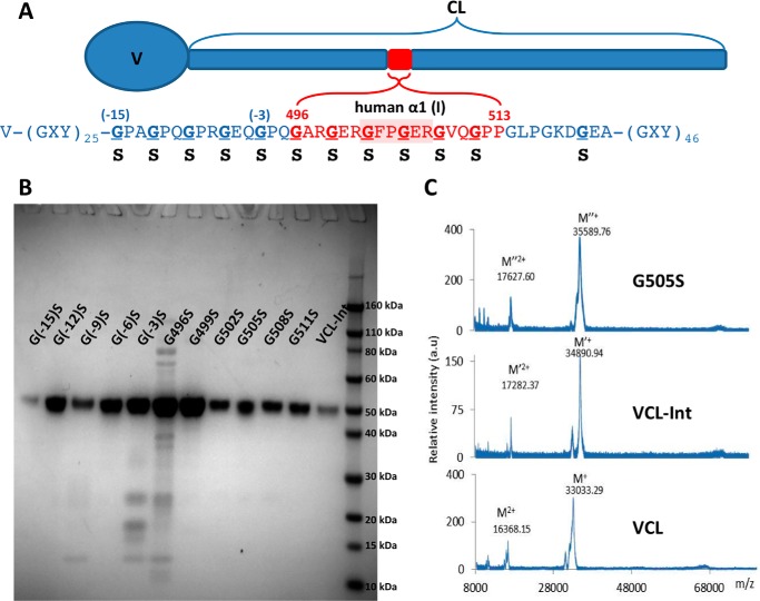
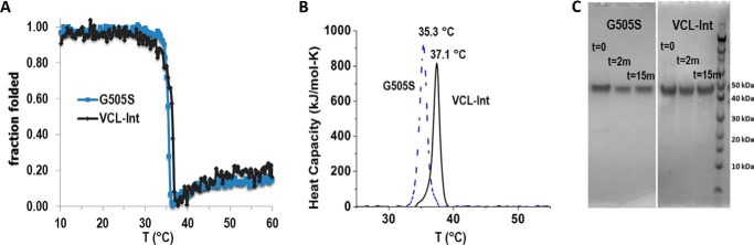
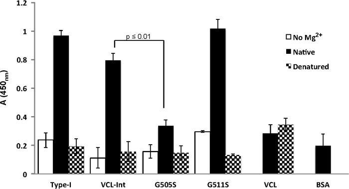
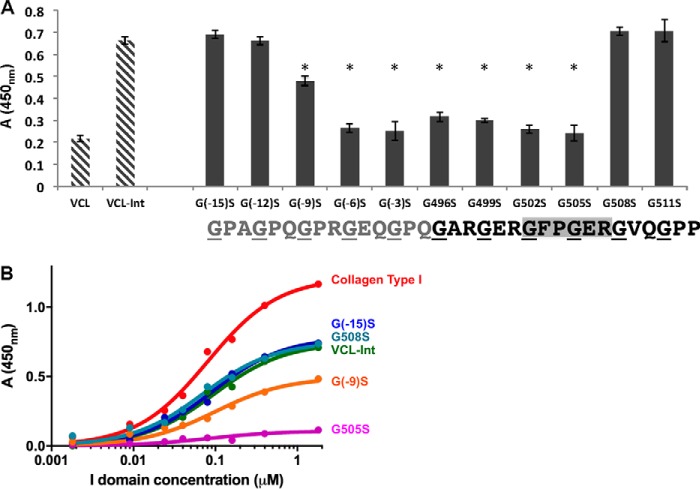
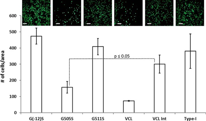
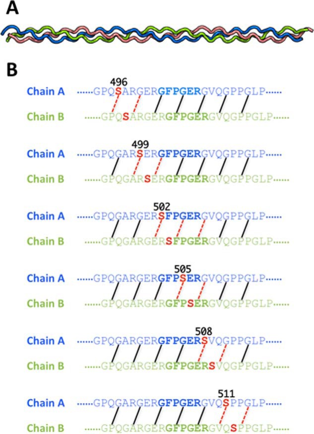
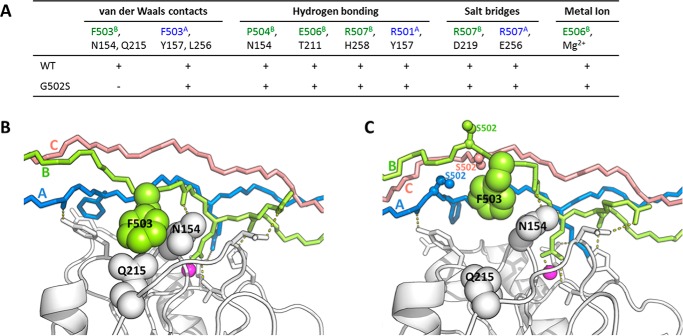
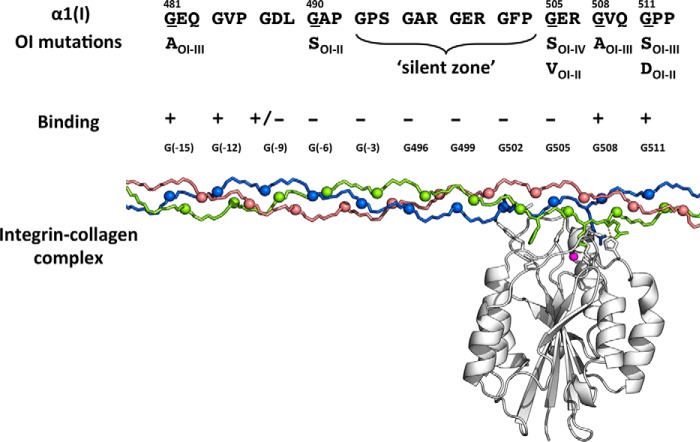
The replacement of one Gly in the essential repeating tripeptide sequence of the type I collagen triple helix results in the dominant hereditary bone disorder osteogenesis imperfecta. The mechanism leading to pathology likely involves misfolding and autophagy, although it has been hypothesized that some mutations interfere with known collagen interactions. Here, the effect of Gly replacements within and nearby the integrin binding GFPGER sequence was investigated using a recombinant bacterial collagen system. When a six-triplet human type I collagen sequence containing GFPGER was introduced into a bacterial collagen-like protein, this chimeric protein bound to integrin. Constructs with Gly to Ser substitutions within and nearby the inserted human sequence still formed a trypsin-resistant triple helix, suggesting a small local conformational perturbation. Gly to Ser mutations within the two Gly residues in the essential GFPGER sequence prevented integrin binding and cell attachment as predicted from molecular dynamics studies of the complex. Replacement of Gly residues C-terminal to GFPGER did not affect integrin binding. In contrast, Gly replacements N-terminal to the GFPGER sequence, up to four triplets away, decreased integrin binding and cell adhesion. This pattern suggests either an involvement of the triplets N-terminal to GFPGER in initial binding or a propagation of the perturbation of the triple helix C-terminal to a mutation site. The asymmetry in biological consequences relative to the mutation site may relate to the observed pattern of osteogenesis imperfecta mutations near the integrin binding site.
Keywords: binding; collagen; extracellular matrix; integrin; missense mutations; molecular dynamics; osteogenesis imperfecta; recombinant protein expression; triple helix.
© 2016 by The American Society for Biochemistry and Molecular Biology, Inc.
Figures
Similar articles
-
Collagen Gly missense mutations: Effect of residue identity on collagen structure and integrin binding.J Struct Biol. 2018 Sep;203(3):255-262. doi: 10.1016/j.jsb.2018.05.003. Epub 2018 May 11. J Struct Biol. 2018. PMID: 29758270 Free PMC article.
-
Molecular underpinnings of integrin binding to collagen-mimetic peptides containing vascular Ehlers-Danlos syndrome-associated substitutions.J Biol Chem. 2019 Sep 27;294(39):14442-14453. doi: 10.1074/jbc.RA119.009685. Epub 2019 Aug 12. J Biol Chem. 2019. PMID: 31406019 Free PMC article.
-
[Effects of Gly mutations N-terminal to the integrin-binding sequence on the structure and function of recombinant collagen].Sheng Wu Gong Cheng Xue Bao. 2025 Apr 25;41(4):1573-1587. doi: 10.13345/j.cjb.240722. Sheng Wu Gong Cheng Xue Bao. 2025. PMID: 40328717 Chinese.
-
Consortium for osteogenesis imperfecta mutations in the helical domain of type I collagen: regions rich in lethal mutations align with collagen binding sites for integrins and proteoglycans.Hum Mutat. 2007 Mar;28(3):209-21. doi: 10.1002/humu.20429. Hum Mutat. 2007. PMID: 17078022 Free PMC article. Review.
-
Nuclear magnetic resonance characterization of peptide models of collagen-folding diseases.Philos Trans R Soc Lond B Biol Sci. 2001 Feb 28;356(1406):159-68. doi: 10.1098/rstb.2000.0761. Philos Trans R Soc Lond B Biol Sci. 2001. PMID: 11260796 Free PMC article. Review.
Cited by
-
Adverse effects of Alport syndrome-related Gly missense mutations on collagen type IV: Insights from molecular simulations and experiments.Biomaterials. 2020 May;240:119857. doi: 10.1016/j.biomaterials.2020.119857. Epub 2020 Feb 12. Biomaterials. 2020. PMID: 32085975 Free PMC article.
-
Collagen Gly missense mutations: Effect of residue identity on collagen structure and integrin binding.J Struct Biol. 2018 Sep;203(3):255-262. doi: 10.1016/j.jsb.2018.05.003. Epub 2018 May 11. J Struct Biol. 2018. PMID: 29758270 Free PMC article.
-
Consequences of Glycine Mutations in the Fibronectin-binding Sequence of Collagen.J Biol Chem. 2016 Dec 30;291(53):27073-27086. doi: 10.1074/jbc.M116.753566. Epub 2016 Oct 31. J Biol Chem. 2016. PMID: 27799304 Free PMC article.
-
Enzymatic Phosphorylation of Ser in a Type I Collagen Peptide.Biophys J. 2018 Dec 18;115(12):2327-2335. doi: 10.1016/j.bpj.2018.11.012. Epub 2018 Nov 16. Biophys J. 2018. PMID: 30527445 Free PMC article.
-
Molecular underpinnings of integrin binding to collagen-mimetic peptides containing vascular Ehlers-Danlos syndrome-associated substitutions.J Biol Chem. 2019 Sep 27;294(39):14442-14453. doi: 10.1074/jbc.RA119.009685. Epub 2019 Aug 12. J Biol Chem. 2019. PMID: 31406019 Free PMC article.
References
Publication types
MeSH terms
Substances
Associated data
- Actions
Grants and funding
LinkOut - more resources
Full Text Sources
Other Literature Sources
