. 2016 Sep;15(9):3030-44.
doi: 10.1074/mcp.M116.060277. Epub 2016 Jul 18.

Defining the Protein-Protein Interaction Network of the Human Protein Tyrosine Phosphatase Family

Affiliations

Defining the Protein-Protein Interaction Network of the Human Protein Tyrosine Phosphatase Family

Xu Li et al. Mol Cell Proteomics. 2016 Sep.

Abstract

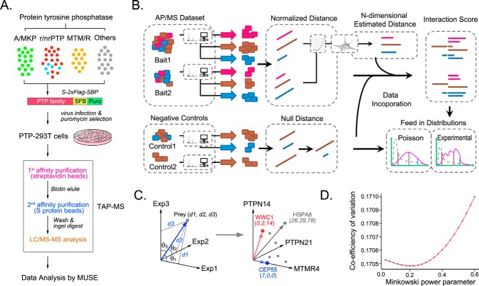
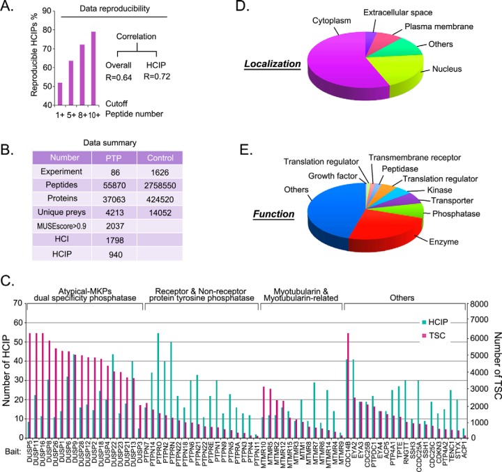
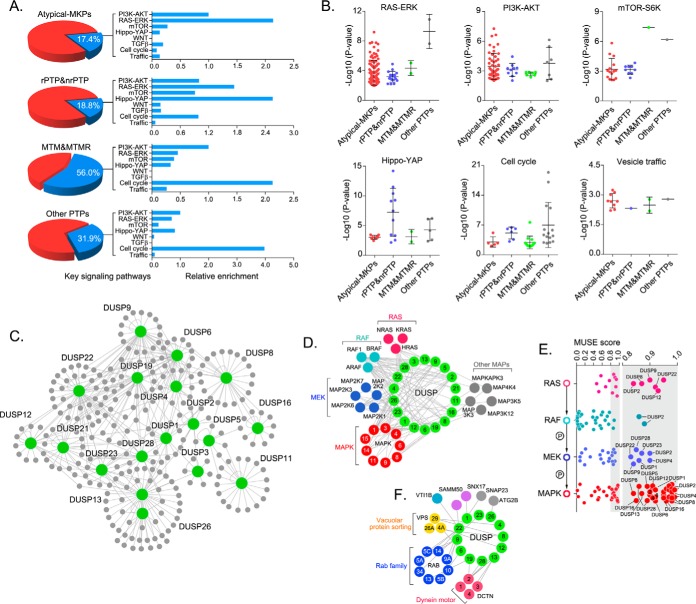
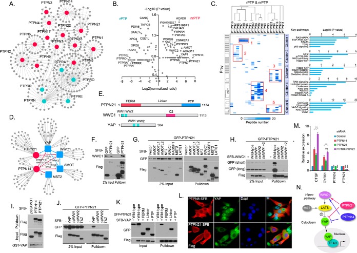
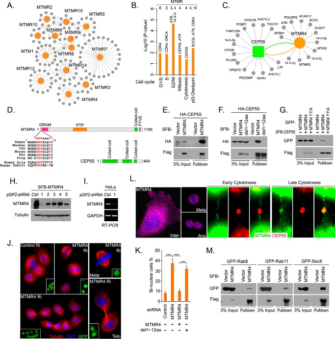
Protein tyrosine phosphorylation, which plays a vital role in a variety of human cellular processes, is coordinated by protein tyrosine kinases and protein tyrosine phosphatases (PTPs). Genomic studies provide compelling evidence that PTPs are frequently mutated in various human cancers, suggesting that they have important roles in tumor suppression. However, the cellular functions and regulatory machineries of most PTPs are still largely unknown. To gain a comprehensive understanding of the protein-protein interaction network of the human PTP family, we performed a global proteomic study. Using a Minkowski distance-based unified scoring environment (MUSE) for the data analysis, we identified 940 high confidence candidate-interacting proteins that comprise the interaction landscape of the human PTP family. Through a gene ontology analysis and functional validations, we connected the PTP family with several key signaling pathways or cellular functions whose associations were previously unclear, such as the RAS-RAF-MEK pathway, the Hippo-YAP pathway, and cytokinesis. Our study provides the first glimpse of a protein interaction network for the human PTP family, linking it to a number of crucial signaling events, and generating a useful resource for future studies of PTPs.


Figures

Fig. 1.
Fig. 1.
Proteomic analysis of the human protein tyrosine phosphatase family. A, Schematic illustration of the major steps of the tandem affinity purification-mass spectrometry (TAP-MS) analysis of the human protein tyrosine phosphatase (PTP) family. Sixty-eight PTP proteins were constructed into a C-terminal SFB-tag fused lentiviral vector using gateway technology. HEK293T cells stably expressing each bait protein were generated by lentiviral infection and puromycin selection. Through standard TAP steps, purified protein complexes were identified by MS analysis, and final interactive proteins were generated by the MUSE statistical model. Three major PTP subfamilies were indicated: atypical-MAPK phosphatase (A/MKP) dual-specificity phosphatases (DSPs), classic PTPs (transmembrane receptor-like phosphatases [rPTPs] and nontransmembrane receptor phosphatases [nrPTPs]), and myotubularin/myotubularin-related (MTM/MTMR) DSPs. B, The workflow of the MUSE algorithm for the TAP-MS data analysis. The diagram depicts the major steps of the MUSE algorithm in the AP/MS data analysis. C–D, Visualization of the example 3-D spaces of TAP-MS and the method of estimating Minkowski power parameter p. An example data set consisting of three independent experiments can be described by a three-dimensional space. In this space, the existence of one prey can be described as one point (C). Exp, experiment. The coefficient of variation of the human PTP data set was evaluated by random drawing and assigning raw spectra counts to random bait-prey combinations, choosing the P that caused minimal system disturbance. The MUSE algorithm simulates the P from 0 to 1, with 0.01 intervals; it then calculates the CV for each preys and combines them to generate the CV for the whole data set (D).
Fig. 2.
Fig. 2.
Summary of the human protein tyrosine phosphatase family proteomics study. A, Data reproducibility for the human PTP family proteomic study. Eighteen atypical-MKP DSP subfamily members were subjected to a biological repeat of the TAP-MS analysis. Overall identified preys and high-confidence candidate interacting proteins (HCIPs) were used to estimate data reproducibility. The bar graph represents raw data reproducibility. The correlation and coefficients were calculated on the basis of raw data. The “cutoff peptide number” meant that we only considered proteins with a certain number of peptides identified. B, The total peptide and protein numbers obtained from the MS analysis are listed. A MUSE score> 0.9 was used as the cutoff to identify HCIPs. C, The total spectral counts (TSCs) and corresponding number of HCIPs for each PTP bait protein are shown together. D, E, Gene ontology annotation for the identified PTP interactors. The cellular localization (D) and cellular functions (E) of the PTP family, based on a GO annotation of their HCIPs, are shown as pie graphs.
Fig. 3.
Fig. 3.
Gene ontology annotation of key signaling pathways and cellular functions of the protein tyrosine phosphatase family. A, B, Identification of key signaling pathways and cellular functions enriched in the protein tyrosine phosphatase (PTP) family. The percentage of identified pathways and functions for each subfamily is indicated (A). The relative enrichment of each pathway and function was compared within each PTP subfamily (A) and between each PTP subfamily (B). Only the statistically significant (p < 0.05) results are shown. C, The interactome of the atypical-MKP dual-specificity phosphatase (DSP) subfamily. D, The cytoscape-generated merged interaction network for the atypical-MKP DSP subfamily and the RAS-ERK pathway. E, The confidence of association between the RAS-ERK pathway and atypical-MKP DSP subfamily was estimated by using th MUSE score. F, The cytoscape-generated merged interaction network for the atypical-MKP DSP subfamily and the identified vesicle trafficking-related proteins.
Fig. 4.
Fig. 4.
Crosstalk between the classic protein tyrosine phosphatase subfamily and the Hippo-YAP pathway. A, Protein interaction network for the classic protein tyrosine phosphatase (PTP) subfamily (transmembrane receptor-like phosphatases [rPTPs] and non-rPTPs [nrPTPs]). rPTP and nrPTP bait proteins were labeled in blue and red, respectively. B, Comparison of rPTP- and nrPTP-associated high-confidence candidate interacting proteins (HCIPs). Overlay of volcano plots of protein enrichments of HCIPs identified in the nrPTP subfamily over the rPTP subfamily, plotted against corresponding p values. The X axis indicates the protein enrichment in the log2 scale, and the Y axis indicates the significance of the changes in the -log10 (p value) scale. X<0 represents the prey enrichment for the rPTP subfamily, and X>0 represents the prey enrichment for the nrPTP subfamily. C, A heatmap was generated from the hierarchical clustering of HCIPs of the classic PTP subfamily. Five prominent HCIP clusters were manually selected, and their signaling pathway annotations are shown. The colors of squares in the heat map represent the number of identified HCIP peptides for each bait protein. D, The merged interaction network among PTPN14, PTPN21, and Hippo pathway components. Reciprocal identification between baits is indicated by a double-headed arrow, and unidirectional identification is indicated by a single-headed arrow. E, Schematic illustration of the domain structures of PTPN21, WWC1, and YAP. F, The association between PTPN21 and WWC1. A pulldown assay was performed with S protein beads, and the indicated proteins were detected by Western blotting. G, PTPN21 specifically interacted with WWC1 in the Hippo pathway. H, Two WW domains of WWC1 were required for its association with PTPN21. I, YAP directly interacted with PTPN14 and PTPN21. Bacterially purified GST-YAP was used for the pulldown experiment. The indicated proteins were detected by Western blot analysis. GST-YAP was shown by Coomassie blue staining. J, Two WW domains of YAP are required for its binding to PTPN21. K, The linker region of PTPN21 mediated its binding to YAP. L, PTPN21 translocated YAP from the nucleus into the cytoplasm. Immunofluorescence staining was performed to detect the localization of endogenous YAP in HeLa cells overexpressing SFB-PTPN5 or SFB-PTPN21. DAPI, nucleus; M, merged. M, Both PTPN14 and PTPN21 suppress YAP activity. Transcripts of YAP target genes were detected by quantitative PCR in HEK293A cells transduced by the indicated shRNAs. ** p < 0.01 and *** p < 0.001. N, A proposed model of Hippo pathway regulation by PTPN14 and PTPN21.
Fig. 5.
Fig. 5.
The roles of the myotubularin/myotubularin-related dual-specificity phosphatase subfamily in cell cycle regulation. A, Protein interaction network of the myotubularin/myotubularin-related (MTM/MTMR) DSP subfamily. B, Identification of the members of the MTM/MTMR DSP subfamily involved in each cell cycle phase. C, The merged protein interaction network between MTMR4 and CEP55. D, Schematic illustration of the domain structures of MTMR4 and CEP55. The GPP(X)3Y motif at the N terminus of MTMR4 is shown, which is conserved in different species. E, The interaction between MTMR4 and CEP55 was validated. Pulldown experiments were performed using S protein beads, and the indicated proteins were detected by Western blotting analysis. F, The N-terminal 12 amino acids of MTMR4 were required for its interaction with CEP55. G, The single site mutation Y11A in the GPP(X)3Y motif of MTMR4 disrupted the interaction between MTMR4 and CEP55. H–I, MTMR4 was down-regulated by shRNA in HeLa cells. MTMR4 protein levels (H) and mRNA levels (I) were detected in HeLa cells transduced with the indicated shRNA. J, Loss of MTMR4 induced a cytokinesis defect. Immunofluorescence staining was performed in shRNA-transduced HeLa cells, as indicated by GFP at different cell cycle phases. Ri, shRNA knockdown; Meta, metaphase; Telo, telophase. K, Loss of MTMR4 induced bi-nuclear cell formation. The percentage of bi-nuclear cells was analyzed for the indicated shRNA and plasmid-transduced cells. *** p < 0.001. L, The vesicle-like localization of MTMR4 in different cell cycle phases. The localizations of SFB-MTMR4 and HA-CEP55 were indicated by immunofluorescence staining. M, MTMR4 associated with endosome proteins, which are involved in membrane fusion at the cleavage stage. HeLa cells were transfected with the indicated plasmids, synchronized at cytokinesis, and subjected to pulldown assays.

Similar articles

Cited by

References

    1. Tonks N. K. (2006) Protein tyrosine phosphatases: from genes, to function, to disease. Nat. Rev. Mol. Cell Biol. 7, 833–846 - PubMed
    1. Zhao S., Sedwick D., and Wang Z. (2014) Genetic alterations of protein tyrosine phosphatases in human cancers. Oncogene 34, 3885–3894 - PMC - PubMed
    1. Ostman A., Hellberg C., and Bohmer F. D. (2006) Protein-tyrosine phosphatases and cancer. Nat. Rev. Cancer 6, 307–320 - PubMed
    1. Alonso A., Sasin J., Bottini N., Friedberg I., Osterman A., Godzik A., Hunter T., Dixon J., and Mustelin T. (2004) Protein tyrosine phosphatases in the human genome. Cell 117, 699–711 - PubMed
    1. Andersen J. N., Jansen P. G., Echwald S. M., Mortensen O. H., Fukada T., Del Vecchio R., Tonks N. K., and Moller N. P. (2004) A genomic perspective on protein tyrosine phosphatases: gene structure, pseudogenes, and genetic disease linkage. FASEB J. 18, 8–30 - PubMed

Publication types

Substances