. 2016 Aug 2;113(31):E4460-6.
doi: 10.1073/pnas.1525730113. Epub 2016 Jul 18.

Directed evolution of a sphingomyelin flippase reveals mechanism of substrate backbone discrimination by a P4-ATPase

Affiliations

Directed evolution of a sphingomyelin flippase reveals mechanism of substrate backbone discrimination by a P4-ATPase

Bartholomew P Roland et al. Proc Natl Acad Sci U S A. .

Abstract

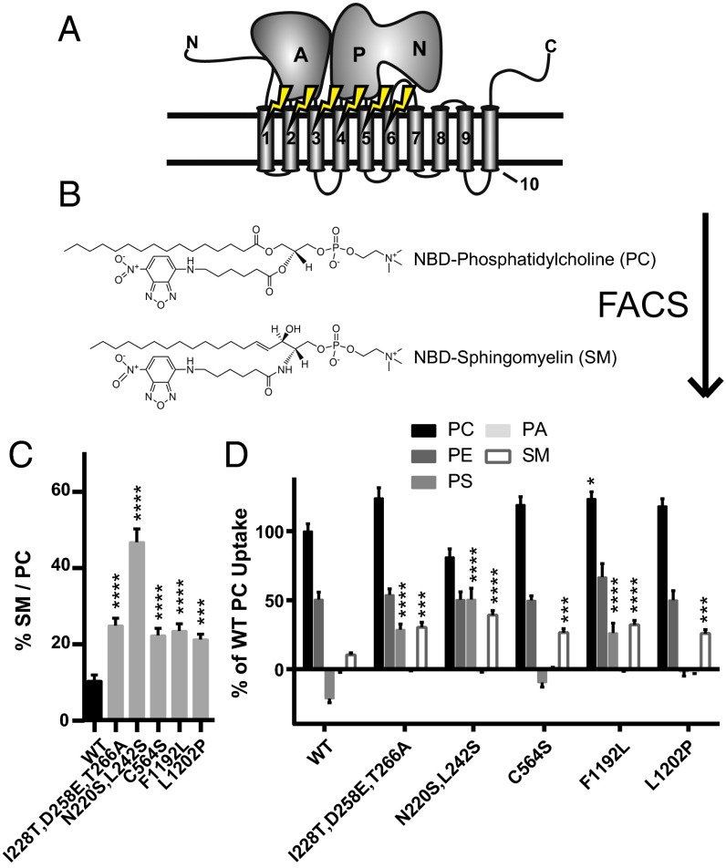
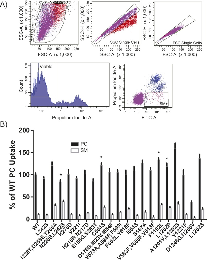
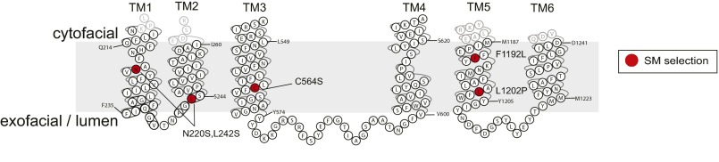
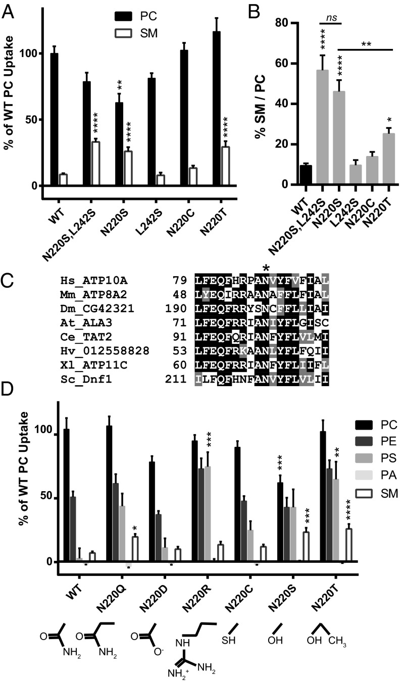
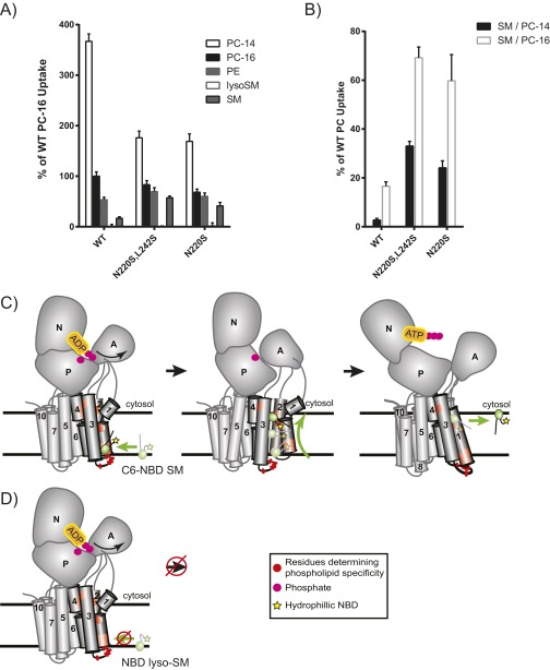
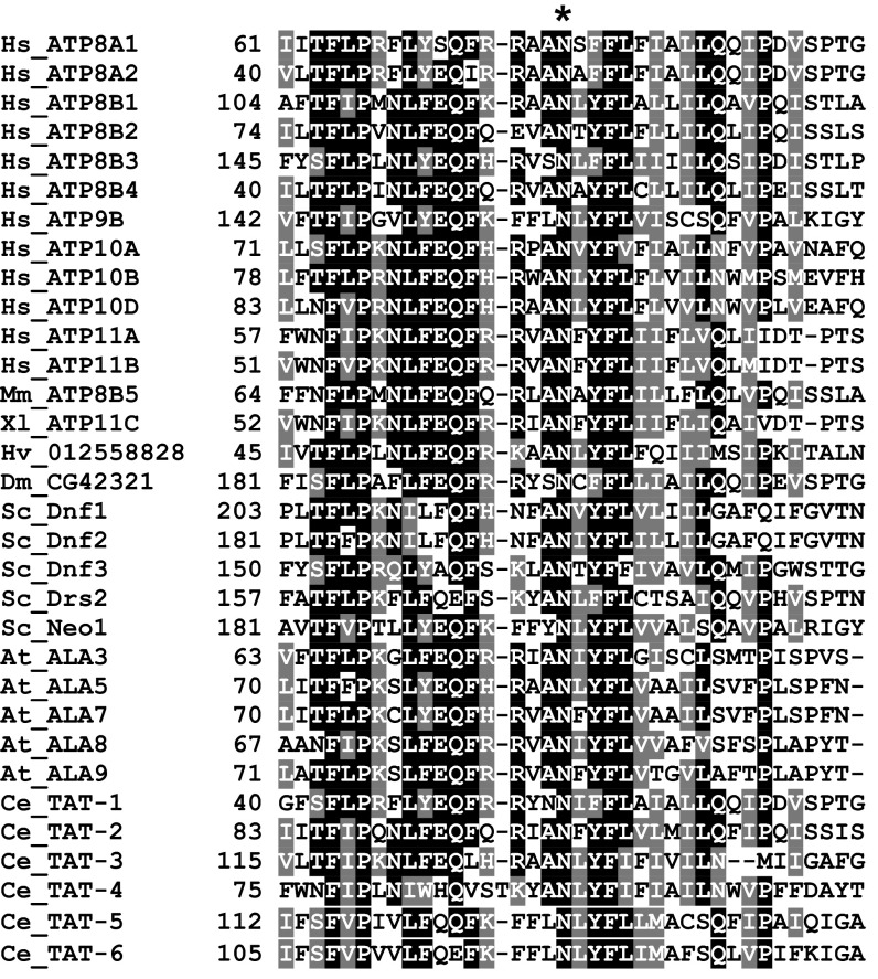
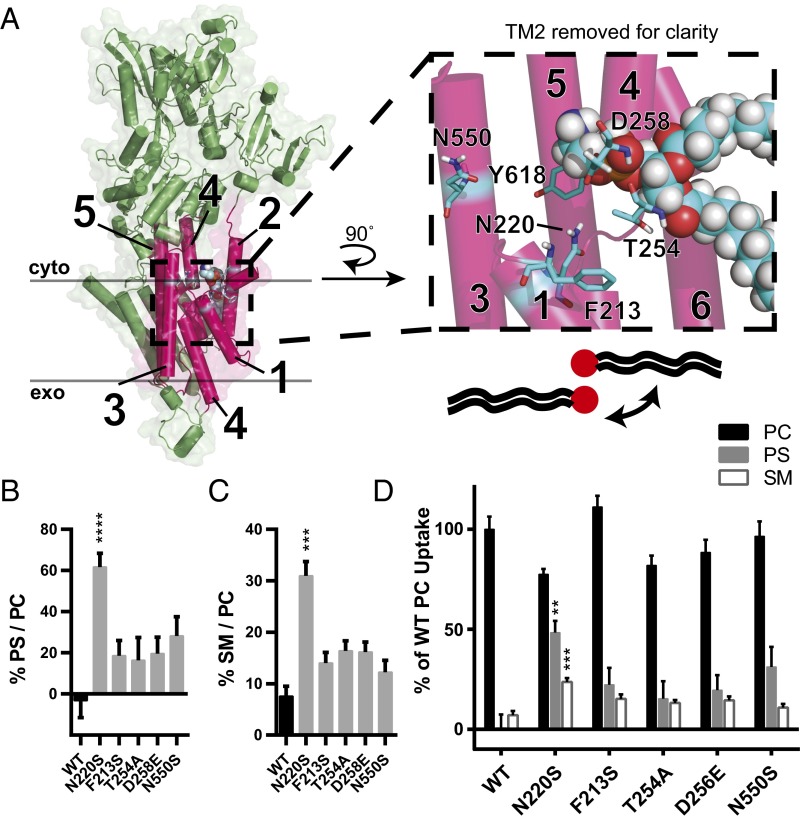
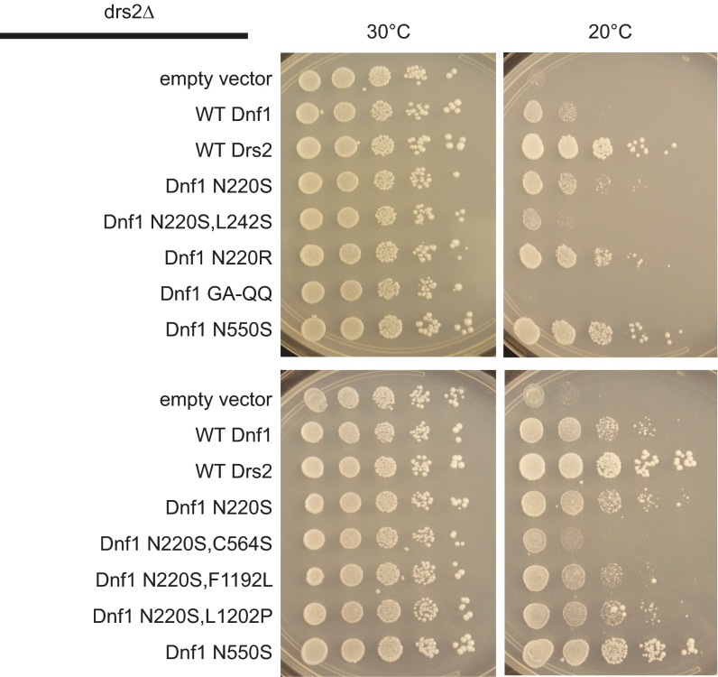
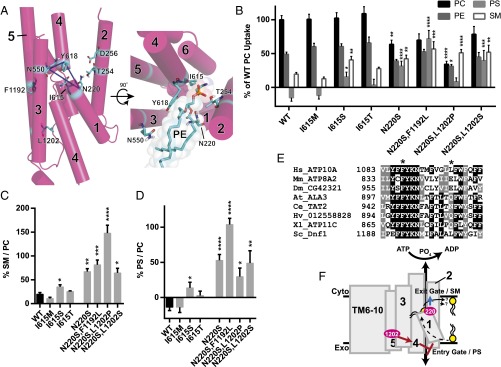
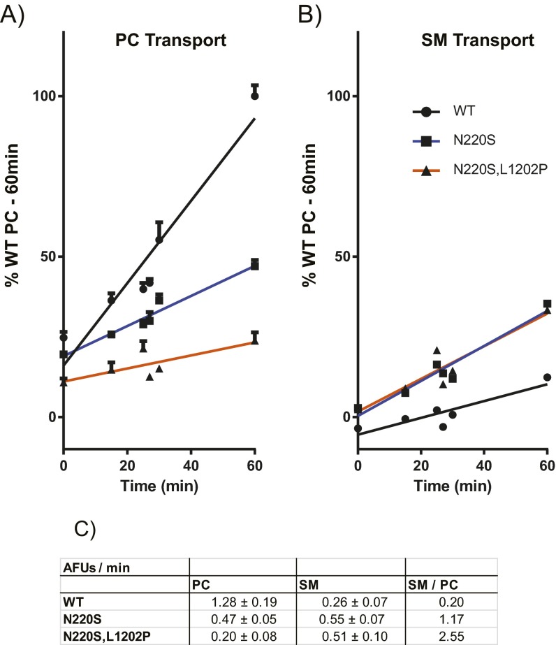
Phospholipid flippases in the type IV P-type ATPase (P4-ATPases) family establish membrane asymmetry and play critical roles in vesicular transport, cell polarity, signal transduction, and neurologic development. All characterized P4-ATPases flip glycerophospholipids across the bilayer to the cytosolic leaflet of the membrane, but how these enzymes distinguish glycerophospholipids from sphingolipids is not known. We used a directed evolution approach to examine the molecular mechanisms through which P4-ATPases discriminate substrate backbone. A mutagenesis screen in the yeast Saccharomyces cerevisiae has identified several gain-of-function mutations in the P4-ATPase Dnf1 that facilitate the transport of a novel lipid substrate, sphingomyelin. We found that a highly conserved asparagine (N220) in the first transmembrane segment is a key enforcer of glycerophospholipid selection, and specific substitutions at this site allow transport of sphingomyelin.

Keywords: P4-ATPase; directed evolution; membrane asymmetry; sphingomyelin.


Conflict of interest statement

The authors declare no conflict of interest.

Figures

Fig. 1.
Fig. 1.
The FACS SM+ screen identifies substitutions throughout TM1, -2, -3, and -5 of Dnf1 that are capable of increasing NBD-SM transport. (A) The first six TM segments were mutagenized by error-prone PCR and selected through FACS for their ability to transport NBD-SM. (B) NBD-SM differs from NBD-PC only in its sphingosine backbone. (C) Five substitutions that double the preference of Dnf1 for NBD-SM were isolated. (D) Measurements of alternative NBD-labeled PLs demonstrate substrate specificity in the SM+ alleles. Negative values occur when the uptake of vector-only control NBD-PL is greater than the uptake of experimental samples. n ≥ 9 ± SEM for all data. Comparisons to WT ratio or substrate uptake were made with one-way ANOVA using Tukey’s post hoc analysis; *P < 0.05; ***P < 0.001; ****P < 0.0001.
Fig. S1.
Fig. S1.
The FACS SM+ screen identifies substitutions through TM1, -2, -3, -4, and -5 that are capable of increasing NBD-SM transport. (A) Dnf1 mutant libraries were gated to select viable, single-cell populations for sorting. Cells containing the top ∼1% of the FITC signal were sorted and cultured. (B) The first-generation hits from the FACS SM+ screen reveal several alleles capable of translocating SM. Several of the SM+ alleles bore substitutions at the same position (e.g., L1202P and L1202S), whereas others were characterized by identical substitutions from different codon changes (e.g., C564S); these observations support previous assertions regarding the complexity of this mutagenesis library (39). n ≥ 9, ± SEM; the asterisk indicates single or double Dnf1 mutants that doubled SM preference relative to WT (see Fig. 1C).
Fig. S2.
Fig. S2.
Topology plot of the top SM-permissive substitutions in TM1–6 of Saccharomyces cerevisiae Dnf1. The cytofacial N220S and F1192L substitutions exhibited increased uptake of NBD-PS, but the exofacial/luminal C564S and L1202P did not influence NBD-PS transport.
Fig. 2.
Fig. 2.
Hydroxyl, but not sulfhydryl, substitutions at the highly conserved N220 are sufficient for SM translocation. (A and B) A serine at position N220 increases NBD-SM selection (A) and preference (B). (C) An alignment of diverse P4-ATPase sequences from mammals to fungi indicates conservation of N220. (D) Substrate transport measurements of alternative biochemical substitutions at position 220 demonstrate the coordination of backbone and headgroup selection. n ≥ 3 for PA examinations, and n ≥ 9 ± SEM for all remaining data. Comparisons to WT ratio or substrate uptake were made with one-way ANOVA using Tukey’s post hoc analysis; *P < 0.05; **P < 0.01; ***P < 0.001; ****P < 0.0001.
Fig. S3.
Fig. S3.
N220S substrate preference is not attributed to differences in substrate solubility or the length of the sn1 acyl position and long-chain base, and is unable to transport NBD-lyso-SM. When measuring SM selection and preference, we were concerned that our observations might be influenced by differences in substrate solubility in NBD-PC and NBD-SM. Differences in solubility would be predicted to alter substrate administration, incorporation into the plasma membrane, and ultimately accessibility to the P4-ATPase. Additionally, the sphingosine backbone carries a trans double bond at position 4. This trans double bond could alter the positioning of the long-chain base within the substrate-binding pocket of the enzyme, thereby influencing the ability to discriminate the length of this base. We measured the uptake of two different NBD-PC substrates with sn1 acyl-chain lengths of 14 and 16 carbons, respectively. Dnf1WT was found to take up PC-14 better than PC-16, as was consistent with its increased solubility. We also tested whether the location of the NBD label on the SM substrate was critical to its transport. Previous work has shown that the yeast Dnf1 enzyme is preferentially a lyso-PL transporter, suggesting that the Dnf1 enzyme recognizes the sn2 C6-NBD GPLs as it would lyso-PLs (38). We examined a lyso-SM substrate with an NBD label at the distal end of the sphingosine chain and found that this substrate could not be recognized by the SM+ mutants. These results likely suggest that the hydrophilic NBD group snorkels up to the aqueous interface of the membrane and perturbs lateral recognition of the lyso-SM substrate by Dnf1. (A) We found that Dnf1N220S,L242S and Dnf1N220S did not enhance the selection of a more soluble NBD-PC containing a shorter sn1 acyl chain but instead reduced the selection of PC-14 similar to that of PC-16. Additionally, the SM+ mutants were unable to recognize NBD-lyso-SM with a fluorophore attached to the distal end of the sphingosine chain. (B) Dnf1N220S,L242S and Dnf1N220S also increased the preference for NBD-SM when normalized to PC-14 and PC-16, suggesting that differences in discriminating substrate solubility or the length of the sn1/long-chain base acyl chain were not responsible for the observed increase in NBD-SM uptake. n ≥ 6 ± SEM. (C) We predict that the C-6 NBD PLs, like the natural substrates, are recognized laterally at the luminal/exofacial aspect of the P4-ATPase between TMs 1, 3, and 4. The PL then is transported through the enzyme and membrane bilayer as a function of ATP catalysis and is released from the P4-ATPase exit gate at the cytofacial aspect of the membrane. (D) Conversely, lyso-SM with the hydrophilic NBD label on the distal end of the long-chain base is predicted to cause the hydrophobic tail to snorkel up to the aqueous interface of the membrane (59), thereby preventing lateral recognition of the substrate at the entry gate.
Fig. S4.
Fig. S4.
A detailed ClustalW sequence alignment around Dnf1 N220. The asterisk demonstrates identity across species and P4-ATPases at this position. Sequence accession numbers are listed in SI Materials and Methods.
Fig. 3.
Fig. 3.
Predicted N220 positioning suggests that substrate backbone and acyl chains are coordinated by TM1 and TM3. (A) Homology model of Dnf1 (PDB ID code 3W5D) with TM 1–6 shown as pink cylinders, the rest of the protein colored green, surface shown, and key residues represented in stick form and colored by element. PE was modeled in the site as crystallized in 3W5D, shown in spheres and colored by element. PM boundaries are indicated. This orientation suggests that the PL headgroup is coordinated by TM2 and TM4, with the substrate backbone and acyl chains protruding between TM2, -4, and -6 (cartoon PL with red headgroup). (Inset) A 90° rotated and enlarged view of the PE site with TM2 removed for clarity. None of the TM2 or TM3 residues alter backbone preference, indicating that the substrate may be oriented in a 180° turn, with the headgroup still between TM2 and TM4 but with the backbone and acyl chains projecting past TM1 and TM3, respectively. (This proposal is illustrated by the cartoon PL and expanded upon with subsequent modeling in Fig. 4A.) (B) Exit gate substitutions F213S, T254A, D258E, and N550S increase PS preference over that of WT. (C) Exit gate substitutions F213S, T254A, D258E, and N550S do not recapitulate the SM preference of N220S. (D) PC, PS, and SM transport of exit gate mutants and N220S. n ≥ 9 ± SEM for all NBD transport data. Comparisons to WT ratio or substrate uptake were made with one-way ANOVA using Tukey’s post hoc analysis; **P < 0.01; ***P < 0.001; ****P < 0.0001.
Fig. S5.
Fig. S5.
Substitutions at the N220 position can partially suppress drs2Δ cold sensitivity, implying that this position cooperates with acyl-chain selection of substrate. Plasmids carrying Dnf1 or Drs2 were transformed into a drs2∆ genetic background and grown at 30 °C or 20 °C. drs2∆ cells have been shown to exhibit growth defects at 20 °C, which can be suppressed by expressing WT Drs2 or Dnf1N550S, a Dnf1 mutation that allows the translocation of diacyl-PS across the membrane. The Dnf1GA-QQ mutation is a gain-of-function substitution that allows lyso-PS transport, but not diacyl-PS, and is unable to suppress the drs2∆ growth inhibition. The Dnf1N220S and Dnf1N220R mutations can transport NBD-PS (Fig. 3) and are weakly capable of altering acyl-chain selection. The compound mutants Dnf1N220S,F1192L and Dnf1N220S,L1202P appear to suppress the cold sensitivity as does WT Dnf1. The figures presented are representative images taken from three independent transformants examined from three growth experiments.
Fig. 4.
Fig. 4.
N220 cooperates with L1202 of TM5 to increase SM selection and preference. (A) Homology model of Dnf1 (3W5D) with TM helices 1–6 shown as pink cylinders and numbered, with key residues represented in stick form and colored by element. Dashed blue lines illustrate the exit gate triad of N220, N550, and Y618; these residues participate in substrate backbone, acyl chain, and headgroup selection, respectively. The rotated view of the model reveals alternative orientation of PL substrate within the exit gate, consistent with the positioning of headgroup-selective, backbone-selective, and acyl chain-selective residues as indicated and shown in sticks. PE is shown in the model as sticks with shaded spheres to illustrate spacing and colored by element. (B) Comparisons of PC, PE, PS, and SM transport among I615M, I615S, I615T, and N220S compound alleles with preferences for SM (C) and PS (D). n ≥ 9 ± SEM for all data. Comparisons to WT ratio or substrate uptake were made with one-way ANOVA using Tukey’s post hoc analysis; *P < 0.05; **P < 0.01; ***P < 0.001; ****P < 0.0001. (E) An alignment of P4-ATPase sequences reveals conservation of F1192 but not L1202. (F) Cartoon schematic of TM-domain substrate transport indicating the putative secondary influence of L1202 for PS selection at the entry gate and the primary selective role of N220 for SM selection at the exit gate.
Fig. S6.
Fig. S6.
Expression and localization of SM+ Dnf1 mutations. N-terminal GFP-tagged Dnf1+, Dnf1N220S, Dnf1N220S,F1192L, and Dnf1N220S,L1202P all display robust PM localization and enrichment at the bud neck. GFP-Dnf1N220S,C564S does not exhibit robust expression or PM/bud neck localization. The representative images were selected from 10 acquisitions of 3 independent transformants. (Scale bar, 5 µm.)
Fig. S7.
Fig. S7.
The compound mutant Dnf1N220S,L1202P increases the specificity of SM transport. (A and B) A time course of NBD-PC (A) and NBD-SM (B) uptake in Dnf1WT, Dnf1N220S, and Dnf1N220S,L1202P. Time points were taken at 0 min (n = 9), 15 min (n = 9), 25 min (n = 3), 27 min (n = 3), 30 min (n = 3), and 60 min (n = 9) ± SEM. Error bars indicate SEM. (C) A table presenting the slope of the linear regression fits in A and B, demonstrating that Dnf1N220S and Dnf1N220S,L1202P have reduced rates of NBD-PC uptake and increased rates of NBD-SM uptake relative to Dnf1WT.

Similar articles

Cited by

References

    1. Vance DE, Vance JE. Biochemistry of Lipids, Lipoproteins and Membranes. 5th Ed Elsevier; Amsterdam: 2008.
    1. van Meer G, Voelker DR, Feigenson GW. Membrane lipids: Where they are and how they behave. Nat Rev Mol Cell Biol. 2008;9(2):112–124. - PMC - PubMed
    1. Devaux PF, Herrmann A. Transmembrane Dynamics of Lipids. Wiley; Hoboken, NJ: 2012.
    1. Fairn GD, et al. High-resolution mapping reveals topologically distinct cellular pools of phosphatidylserine. J Cell Biol. 2011;194(2):257–275. - PMC - PubMed
    1. Seigneuret M, Devaux PF. ATP-dependent asymmetric distribution of spin-labeled phospholipids in the erythrocyte membrane: Relation to shape changes. Proc Natl Acad Sci USA. 1984;81(12):3751–3755. - PMC - PubMed

Publication types

MeSH terms

LinkOut - more resources