. 2016 Aug 16;113(33):9155-61.
doi: 10.1073/pnas.1605146113. Epub 2016 Jul 18.

Extracellular vesicles and viruses: Are they close relatives?

Affiliations

Extracellular vesicles and viruses: Are they close relatives?

Esther Nolte-'t Hoen et al. Proc Natl Acad Sci U S A. .

Abstract

Extracellular vesicles (EVs) released by various cells are small phospholipid membrane-enclosed entities that can carry miRNA. They are now central to research in many fields of biology because they seem to constitute a new system of cell-cell communication. Physical and chemical characteristics of many EVs, as well as their biogenesis pathways, resemble those of retroviruses. Moreover, EVs generated by virus-infected cells can incorporate viral proteins and fragments of viral RNA, being thus indistinguishable from defective (noninfectious) retroviruses. EVs, depending on the proteins and genetic material incorporated in them, play a significant role in viral infection, both facilitating and suppressing it. Deciphering the mechanisms of EV-cell interactions may facilitate the design of EVs that inhibit viral infection and can be used as vehicles for targeted drug delivery.

Keywords: defective viruses; exosomes; extracellular vesicles; infection; viruses.


Conflict of interest statement

The authors declare no conflict of interest.

Figures

Fig. 1.
Fig. 1.
Structural similarities between EVs and virions. Cells infected with enveloped RNA (retro)viruses release vesicles containing a variety of host and viral factors. On one extreme, there are EVs consisting entirely of host cell components (blue), and on the other extreme there are infectious viruses surrounded by a host lipid bilayer and containing all of the virus-specific molecules (red) necessary for infectivity. In virus-infected cells, EVs incorporate fragments of the viral genome and viral (glyco)proteins. Moreover, virus infections modify the incorporation of host proteins and RNAs into EVs (light blue). Such infection-induced EVs and the so-called defective viruses and virus-like particles are intermediate entities, and the border between them seems not to exist.
Fig. 2.
Fig. 2.
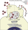
Similarities between biogenesis of EVs and virions. EVs and enveloped retrovirus particles (e.g., HIV) are simultaneously released by infected cells and share pathways for biogenesis at the plasma membrane or at multivesicular bodies (MVBs). For example, proteins of the ESCRT complex and tetraspanins are involved in both virion and EV formation. Viral RNA (red) enters the cytoplasm, after which Gag-mediated virion assembly takes place in the MVB or at the plasma membrane. MVB can contain both virions and EVs and are released from the cell after fusion of the MVB with the plasma membrane through the action of Rab, SNARE, and SNAP proteins. Defective viruses are also formed but are noninfectious because of the lack of essential viral components. Whereas specific host proteins and RNAs (blue), such as CD63 and APOBEC3G, can be incorporated into virions, viral components (red) are also incorporated in the plethora of EV types released by the cells. These include fragments of the viral genome, viral miRNAs, and viral (glyco)proteins, such as Nef and Gag. This intertwining of their pathways for biogenesis blurs the distinction between virions and EVs.
Fig. 3.
Fig. 3.
Proviral and antiviral effects of EVs released by retrovirus-infected cells. Retrovirus infection can lead to the release of modified EVs that either facilitate or suppress infection. Potential antiviral effects include (A) EV-mediated delivery of antiviral components, such as APOBEC3G, to increase resistance to infection; (B) spread of TLR ligands, such as viral RNA, via EVs to warn nonsusceptible neighboring cells of the presence of viral infection; and (C) provision of antigen presenting cells with viral antigen to facilitate the initiation of adaptive immune responses. Potential proviral effects include (D) inhibition of the neutralizing effect of EV, leading to decreased binding of EV to virions and an increase in the number of virions that may infect other cells; (E) EV-mediated delivery of viral components (e.g., Nef) that induce induce cell senescence or death of antiviral immune cells; (F) EV-mediated delivery of viral components that suppress the function of immune cells (e.g., Nef-induced down-regulation of antibody production by B cells); and (G) increase of the pool of virus-susceptible cells, e.g., by transference of coreceptors for virus binding to other cells.

Similar articles

Cited by

References

    1. Colombo M, Raposo G, Théry C. Biogenesis, secretion, and intercellular interactions of exosomes and other extracellular vesicles. Annu Rev Cell Dev Biol. 2014;30:255–289. - PubMed
    1. Yáñez-Mó M, et al. Biological properties of extracellular vesicles and their physiological functions. J Extracell Vesicles. 2015;4:27066. - PMC - PubMed
    1. Valadi H, et al. Exosome-mediated transfer of mRNAs and microRNAs is a novel mechanism of genetic exchange between cells. Nat Cell Biol. 2007;9(6):654–659. - PubMed
    1. Chahar HS, Bao X, Casola A. Exosomes and their role in the life cycle and pathogenesis of RNA viruses. Viruses. 2015;7(6):3204–3225. - PMC - PubMed
    1. Feng Z, et al. A pathogenic picornavirus acquires an envelope by hijacking cellular membranes. Nature. 2013;496(7445):367–371. - PMC - PubMed

Publication types