

Interleukin-29 Enhances Synovial Inflammation and Cartilage Degradation in Osteoarthritis
- PMID: 27433031
- PMCID: PMC4940582
- DOI: 10.1155/2016/9631510
Interleukin-29 Enhances Synovial Inflammation and Cartilage Degradation in Osteoarthritis
Abstract
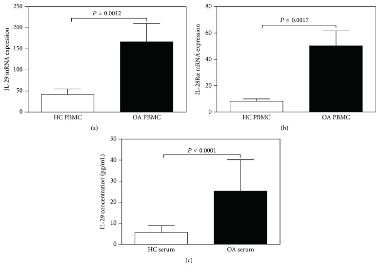
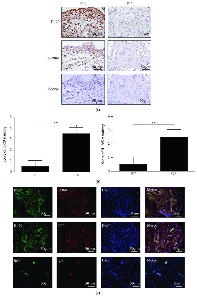
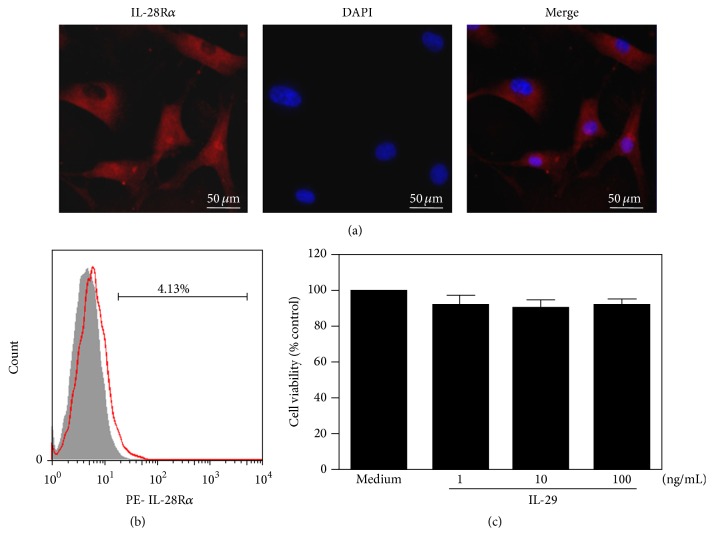
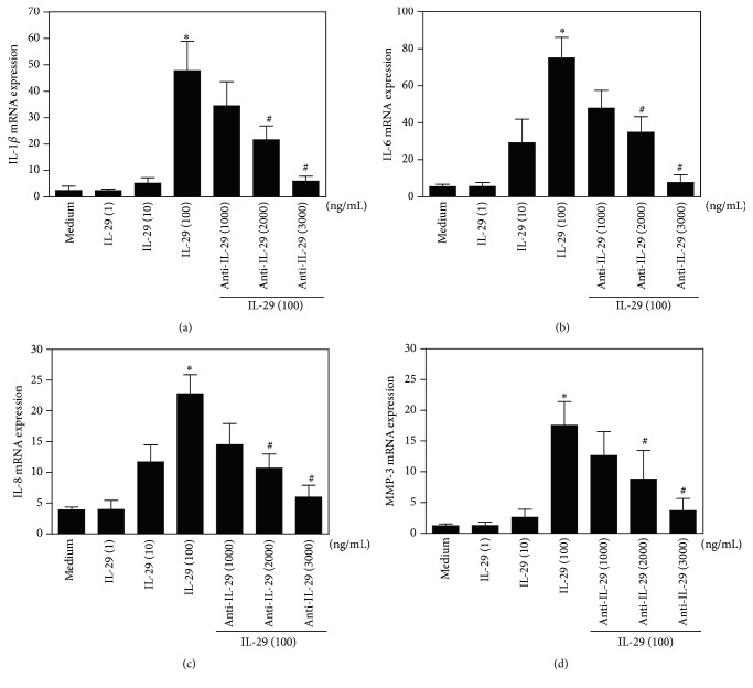
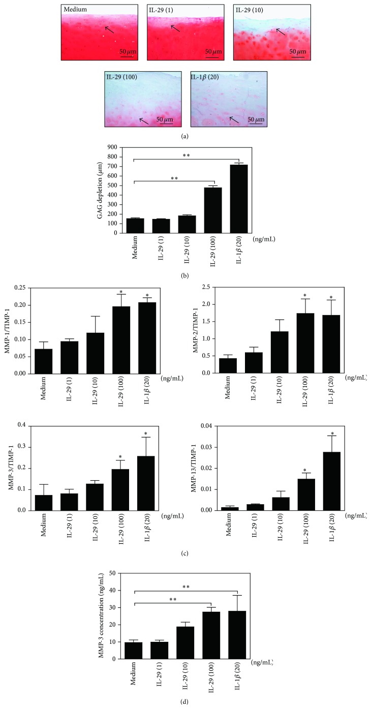
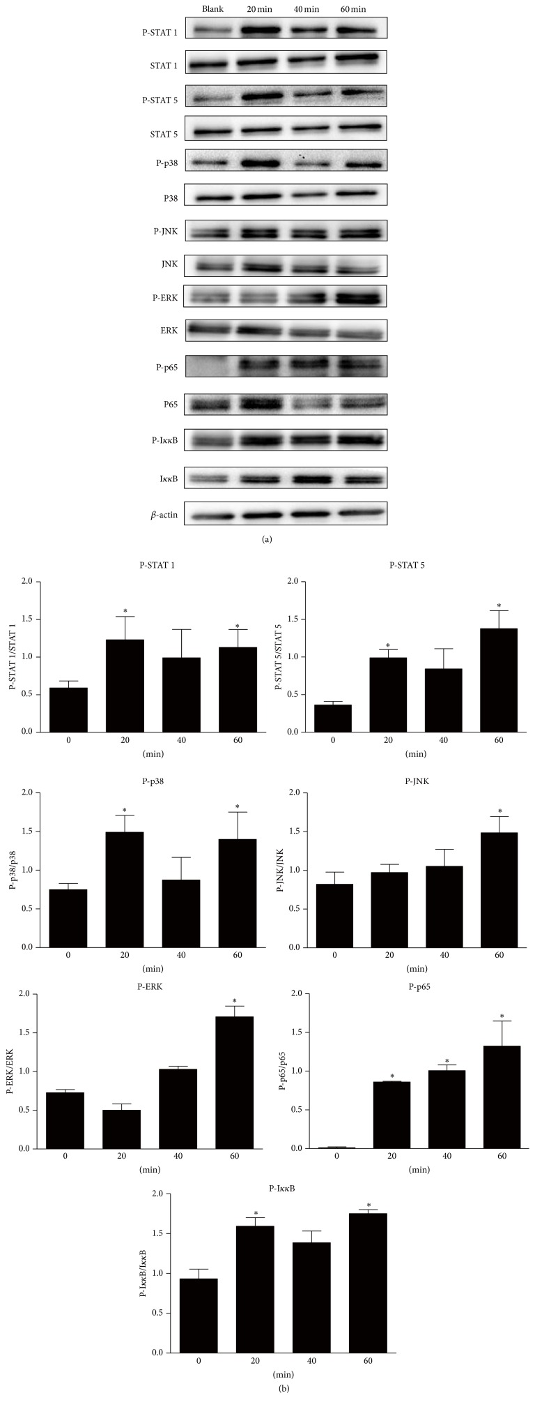
We have recently shown that IL-29 was an important proinflammatory cytokine in pathogenesis of rheumatoid arthritis (RA). Inflammation also contributes to the pathogenesis of osteoarthritis (OA). The aim of this study was to investigate the effect and mechanism of IL-29 on cytokine production and cartilage degradation in OA. The mRNA levels of IL-29 and its specific receptor IL-28Ra in peripheral blood mononuclear cells (PBMCs) were significantly increased in OA patients when compared to healthy controls (HC). In the serum, IL-29 protein levels were higher in OA patients than those in HC. Immunohistochemistry revealed that both IL-29 and IL-28Ra were dramatically elevated in OA synovium compared to HC; synovial fibroblasts (FLS) and macrophages were the main IL-29-producing cells in OA synovium. Furthermore, recombinant IL-29 augmented the mRNA expression of IL-1β, IL-6, IL-8, and matrix-metalloproteinase-3 (MMP-3) in OA FLS and increased cartilage degradation when ex vivo OA cartilage explant was coincubated with OA FLS. Finally, in OA FLS, IL-29 dominantly activated MAPK and nuclear factor-κB (NF-κB), but not Jak-STAT and AKT signaling pathway as examined by western blot. In conclusion, IL-29 stimulates inflammation and cartilage degradation by OA FLS, indicating that this cytokine is likely involved in the pathogenesis of OA.
Figures
Similar articles
-
Interleukin-29 modulates proinflammatory cytokine production in synovial inflammation of rheumatoid arthritis.Arthritis Res Ther. 2012 Oct 19;14(5):R228. doi: 10.1186/ar4067. Arthritis Res Ther. 2012. PMID: 23078630 Free PMC article.
-
Upregulated expression of CCR3 in osteoarthritis and CCR3 mediated activation of fibroblast-like synoviocytes.Cytokine. 2016 Jan;77:211-9. doi: 10.1016/j.cyto.2015.09.012. Epub 2015 Sep 26. Cytokine. 2016. PMID: 26409848
-
[An increased level of interleukin 27 in peripheral blood mononuclear cells and fibroblasts like synoviocytes of patients with rheumatoid arthritis or osteoarthritis].Xi Bao Yu Fen Zi Mian Yi Xue Za Zhi. 2015 Dec;31(12):1673-6. Xi Bao Yu Fen Zi Mian Yi Xue Za Zhi. 2015. PMID: 26648303 Chinese.
-
The role of cytokines in osteoarthritis pathophysiology.Biorheology. 2002;39(1-2):237-46. Biorheology. 2002. PMID: 12082286 Review.
-
The roles of catabolic factors in the development of osteoarthritis.Tissue Eng Part B Rev. 2014 Aug;20(4):355-63. doi: 10.1089/ten.TEB.2013.0377. Epub 2013 Dec 11. Tissue Eng Part B Rev. 2014. PMID: 24172137 Free PMC article. Review.
Cited by
-
The protective effects of Olmesartan against interleukin-29 (IL-29)-induced type 2 collagen degradation in human chondrocytes.Bioengineered. 2022 Jan;13(1):1802-1813. doi: 10.1080/21655979.2021.1997090. Bioengineered. 2022. Retraction in: Bioengineered. 2024 Dec;15(1):2299551. doi: 10.1080/21655979.2024.2299551. PMID: 35012432 Free PMC article. Retracted.
-
Type III Interferons: Emerging Roles in Autoimmunity.Front Immunol. 2021 Nov 26;12:764062. doi: 10.3389/fimmu.2021.764062. eCollection 2021. Front Immunol. 2021. PMID: 34899712 Free PMC article. Review.
-
Long non-coding RNA DANCR regulates proliferation and apoptosis of chondrocytes in osteoarthritis via miR-216a-5p-JAK2-STAT3 axis.Biosci Rep. 2018 Dec 14;38(6):BSR20181228. doi: 10.1042/BSR20181228. Print 2018 Dec 21. Biosci Rep. 2018. PMID: 30361290 Free PMC article.
-
Evaluation of IL-29 in Euthyroid Patients with Graves' Orbitopathy: A Preliminary Study.Mediators Inflamm. 2020 Jul 9;2020:4748612. doi: 10.1155/2020/4748612. eCollection 2020. Mediators Inflamm. 2020. PMID: 32694926 Free PMC article.
-
miR-373 regulates inflammatory cytokine-mediated chondrocyte proliferation in osteoarthritis by targeting the P2X7 receptor.FEBS Open Bio. 2018 Feb 10;8(3):325-331. doi: 10.1002/2211-5463.12345. eCollection 2018 Mar. FEBS Open Bio. 2018. PMID: 29511609 Free PMC article.
References
MeSH terms
Substances
LinkOut - more resources
Full Text Sources
Other Literature Sources
Medical
Miscellaneous
