Purtscher-like retinopathy in acute alcoholic pancreatitis
- PMID: 27433040
- PMCID: PMC4932793
- DOI: 10.4103/0974-620X.184531
Purtscher-like retinopathy in acute alcoholic pancreatitis
Abstract
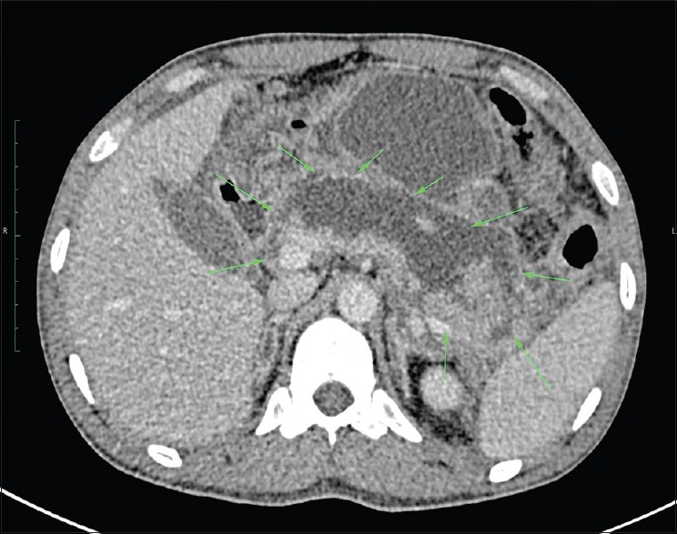
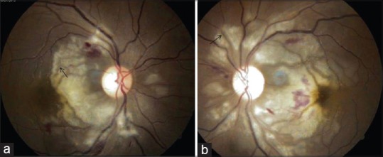
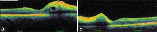
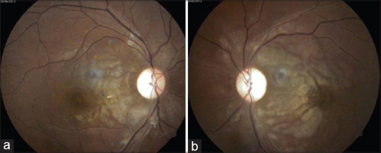
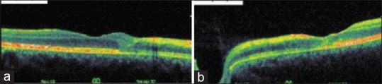
A 23-year-old man with a history of alcoholism presented with vomiting, fever, and sharp epigastric pain radiating to the back and flanks. He was diagnosed as a case of acute alcoholic pancreatitis on the basis of clinical findings and investigations. On the next day of presentation, he developed sudden bilateral visual loss. His best-corrected visual acuity was finger counting at one-foot distance in both eyes. He had diffuse whitening in the circumpapillary area, haloes around the retinal vessels (Purtscher flecken) and intra-retinal hemorrhages on ophthalmoscopic examination. Optical coherence tomography revealed bilateral macular edema. These findings were characteristic of Purtscher-like retinopathy. The patient showed systemic and visual improvement at 8 weeks follow-up after receiving the conventional treatment for acute alcoholic pancreatitis. This case emphasizes the importance of fundus examination by an ophthalmologist in the diagnosis of this rare under-diagnosed entity.
Keywords: Cotton-wool spots; Purtscher flecken; optical coherence tomography.
Figures
Similar articles
-
Purtscher's and Purtscher-like retinopathy etiology, features, management, and outcomes: A summative systematic review of 168 cases.PLoS One. 2024 Sep 6;19(9):e0306473. doi: 10.1371/journal.pone.0306473. eCollection 2024. PLoS One. 2024. PMID: 39240905 Free PMC article.
-
Internal limiting membrane separation and posterior vitreous hyperreflective dots: novel OCT findings in Purtscher-like retinopathy.BMC Ophthalmol. 2024 Mar 27;24(1):137. doi: 10.1186/s12886-024-03413-w. BMC Ophthalmol. 2024. PMID: 38532407 Free PMC article.
-
Purtscher-like retinopathy following valsalva maneuver effect: case report.J Med Case Rep. 2011 Aug 1;5:338. doi: 10.1186/1752-1947-5-338. J Med Case Rep. 2011. PMID: 21806816 Free PMC article.
-
Purtscher-like retinopathy following coronary artery bypass grafting in an antiphospholipid syndrome patient: a case report.BMC Ophthalmol. 2023 May 4;23(1):197. doi: 10.1186/s12886-023-02935-z. BMC Ophthalmol. 2023. PMID: 37142991 Free PMC article.
-
[Purtscher-like retinopathy associated with antibiotic anaphylaxis].Nan Fang Yi Ke Da Xue Xue Bao. 2018 Mar 20;38(3):239-242. doi: 10.3969/j.issn.1673-4254.2018.03.01. Nan Fang Yi Ke Da Xue Xue Bao. 2018. PMID: 29643027 Free PMC article. Review.
Cited by
-
Purtscher-like Retinopathy in a Patient with Acute Pancreatitis: A Case Report and Literature Review.Brown J Hosp Med. 2023 Apr 1;2(2):73885. doi: 10.56305/001c.73885. eCollection 2023. Brown J Hosp Med. 2023. PMID: 40046155 Free PMC article.
-
Purtscher's and Purtscher-like retinopathy etiology, features, management, and outcomes: A summative systematic review of 168 cases.PLoS One. 2024 Sep 6;19(9):e0306473. doi: 10.1371/journal.pone.0306473. eCollection 2024. PLoS One. 2024. PMID: 39240905 Free PMC article.
-
Purtscher's-like retinopathy as a rare complication of acute alcoholic pancreatitis.Prz Gastroenterol. 2021;16(2):170-173. doi: 10.5114/pg.2021.106669. Epub 2021 Jun 4. Prz Gastroenterol. 2021. PMID: 34276846 Free PMC article. No abstract available.
References
-
- Purtscher O. Noch unbekannte befunde nach schadeltrauma. Ber Dtsch Ophthalmol Ges. 1910;36:294–310.
-
- Behrens-Baumann W, Scheurer G, Schroer H. Pathogenesis of purtscher's retinopathy. An experimental study. Graefes Arch Clin Exp Ophthalmol. 1992;230:286–91. - PubMed
-
- Inkeles DM, Walsh JB. Retinal fat emboli as sequela to acute pancreatitis. Am J Ophthalmol. 1975;80:935–8. - PubMed
Publication types
LinkOut - more resources
Full Text Sources
Other Literature Sources