Review
. 2014 Nov;22(4):205-12.
doi: 10.1177/1742271X14545911. Epub 2014 Jul 30.

Can ultrasound help to manage patients with scrotal trauma?

Affiliations
Review

Can ultrasound help to manage patients with scrotal trauma?

T Adlan et al. Ultrasound. 2014 Nov.

Abstract

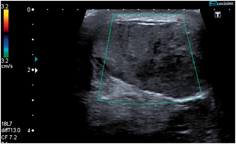
Traumatic injuries to the scrotum are uncommon but, when they do occur, frequently lead to serious complications. Early complications include testicular infarction, necrosis and abscess formation; in the longer-term trauma may result in testicular atrophy and subfertility. Early surgical intervention in patients with testicular rupture can significantly improve the clinical outcome and reduce the need for delayed orchidectomy. However, clinical examination of the scrotum following trauma is difficult and frequently inaccurate; this may result in incorrect triage of patients for surgical exploration. Scrotal ultrasound can reliably assess scrotal injuries and diagnose testicular rupture with a high level of accuracy. Additionally, ultrasound can provide important information regarding testicular perfusion, which can further inform decisions on surgical management. This article reviews the sonographic findings that may be encountered in patients with scrotal trauma, with an emphasis on blunt trauma. It describes the pivotal role that ultrasound can play in the accurate triage of these patients to surgical or conservative management.

Keywords: Ultrasound testis; testicular rupture; testicular trauma; ultrasound scrotum.


Figures

Figure 1
Figure 1
Testicular rupture. A nodule of testicular parenchyma is seen deforming the normally smooth testicular surface (arrows). There is a moderate haematocoele present
Figure 2
Figure 2
Testicular rupture. In this patient there is an abnormal irregular contour of the anterior surface of the lower pole of testis (arrows), a haematocoele is present
Figure 3
Figure 3
Testicular rupture. Large areas of low echogenicity are present within the testicular parenchyma indicating areas of testicular haematoma/contusion (arrows). There is a large extra-testicular haematoma (H)
Figure 4
Figure 4
Testicular rupture. In this case no intra-testicular blood flow can be demonstrated with power Doppler. It is probable that this testis is completely devascularised and orchidectomy is likely to be necessary
Figure 5
Figure 5
Testicular fracture. An irregular vertical fracture line is seen through the lower pole of the testis (arrows) indicating a testicular fracture line
Figure 6
Figure 6
Intra-testicular haematoma. There is a moderately sized irregular echo-poor haematoma within the upper pole of the testis. The remaining testicular parenchyma was well perfused and therefore this patient was managed conservatively. Follow-up scans would be required in this situation to document resolution of the haematoma, detect developing complications and exclude an underlying testicular tumour
Figure 7
Figure 7
Extra-testicular haematoma. There is a large echogenic haematoma indenting and compressing the testis. Surgical evacuation should be considered in this situation if there is evidence of reduced testicular perfusion
Figure 8
Figure 8
Large post traumatic haematocoele. This is a follow-up scan two weeks after scrotal trauma showing that the haematocoele has become heavily septated
Figure 9
Figure 9
Traumatic testicular torsion. This 17-year-old patient presented one week following a kick in the scrotum with increasing scrotal pain and swelling. There is no detectable flow within the testis, areas of low echogenicity within the testis indicate the presence of testicular infarction. Surgical exploration confirmed testicular torsion, the testis was non-viable and orchidectomy was performed
Figure 10
Figure 10
Testicular infarction following hernia repair. This patient presented 10 days’ post-inguinal hernia repair with scrotal swelling and pain. Contrast enhanced ultrasound (split screen image to the readers left) shows that the testis is almost entirely avascular with only a small nodule of enhancing parenchyma at the upper pole (arrow)

References

    1. Munter DW, Faleski EJ. Blunt scrotal trauma: emergency department evaluation and management. Am J Emerg Med 1989; 7: 227–34. - PubMed
    1. Cross JJL, Berman LH, Elliott PG, Irving S. Scrotal trauma: a cause of testicular atrophy. Clin Radiol 1999; 54: 317–20. - PubMed
    1. Kukadia AN, Ercole CJ, Gleich P, Hensleigh H, Pryor JL. Testicular trauma: potential impact on reproductive function. J Urol 1996; 156: 1643–6. - PubMed
    1. Lin WW, Kim ED, Quesada ET, Lipshultz LI, Coburn M. Unilateral testicular injury from external trauma: evaluation of semen quality and endocrine parameters. J Urol 1998; 159: 841–3. - PubMed
    1. Chandra RV, Dowling RJ, Ulubasoglu M, Haxhimolla H, Costello AJ. Rational approach to diagnosis and management of blunt scrotal trauma. Urology 2007; 70: 230–4. - PubMed

LinkOut - more resources