

Solitary fibrous tumour of the cheek: An unusual presentation of a rare soft tissue tumour
- PMID: 27433225
- PMCID: PMC4760550
- DOI: 10.1177/1742271X14554145
Solitary fibrous tumour of the cheek: An unusual presentation of a rare soft tissue tumour
Abstract
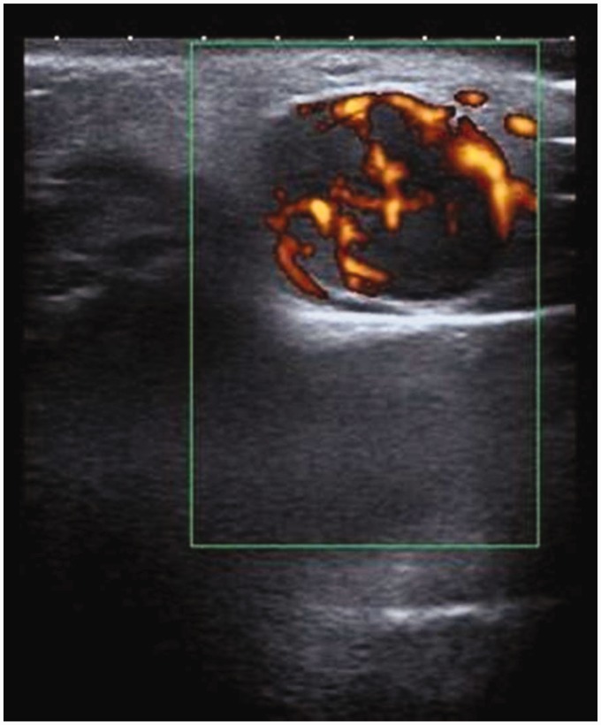
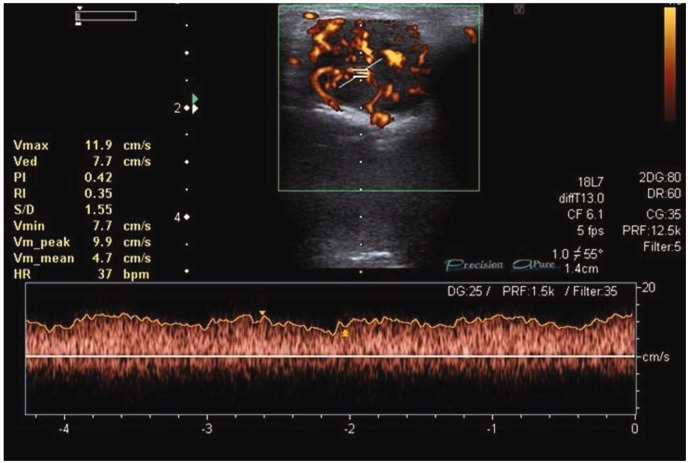
This case report discusses the unusual presentation and ultrasound features of a solitary fibrous tumour of the face. Solitary fibrous tumour is an uncommon form of soft tissue tumour which, although seen predominantly within the lung pleura, can occur throughout the body in sites such as the peritoneum, mediastinum and head and neck. Ultrasound is an excellent imaging modality in the assessment of soft tissue masses in the head and neck. The ultrasound features demonstrated by this example of solitary fibrous tumour are reviewed. This report also highlights that ultrasound alone is ultimately limited in reaching a definitive diagnosis. The roles of other investigations such as ultrasound-guided biopsy and cross-sectional imaging are discussed.
Keywords: Diagnostic imaging; head and neck; tumour; ultrasound; ultrasound appearances.
Figures


Similar articles
-
Solitary fibrous tumour of the face: a rare case report.Br J Plast Surg. 2002 Jan;55(1):75-7. doi: 10.1054/bjps.2001.3723. Br J Plast Surg. 2002. PMID: 11783975 Review.
-
Solitary fibrous tumour of the face: a rare case report.J Plast Reconstr Aesthet Surg. 2010 Jan;63(1):e13-5. doi: 10.1016/j.bjps.2009.05.026. Epub 2009 Jun 13. J Plast Reconstr Aesthet Surg. 2010. PMID: 19527945
-
Solitary fibrous tumour of the soft tissue of the face: a case report.B-ENT. 2006;2(4):201-4. B-ENT. 2006. PMID: 17256410
-
Inaccuracy of fine-needle biopsy in the diagnosis of solitary fibrous tumour of the liver.Asian J Surg. 2008 Oct;31(4):195-8. doi: 10.1016/S1015-9584(08)60085-8. Asian J Surg. 2008. PMID: 19010762
-
Sinonasal and rhinopharyngeal solitary fibrous tumour: a case report and review of the literature.Acta Otorhinolaryngol Ital. 2015 Dec;35(6):455-8. doi: 10.14639/0392-100X-163813. Acta Otorhinolaryngol Ital. 2015. PMID: 26900253 Free PMC article. Review.
References
-
- Klemperer P, Rabin CB. Primary neoplasms of the pleura: a report of 5 cases. Arch Pathol 1931; 11: 385–412.
-
- Witkin GB, Rosai J. Solitary fibrous tumour of the upper respiratory tract. A report of six cases. Am J Surg Pathol 1989; 13: 547–57. - PubMed
-
- Barnes L, Eveson JW, Reichart P, et al. World Health Organization Classification of Tumours. Pathology and Genetics of Head and Neck Tumours, Lyon: IARC Press, 2005.
-
- Papadakis I, Koudounarakis E, Haniotis V, et al. Atypical solitary fibrous tumour of the nose and maxillary sinus. Head Neck 2013; 35: E77–9. - PubMed
Publication types
LinkOut - more resources
Full Text Sources
Other Literature Sources