Review
. 2015 Aug;23(3):158-65.
doi: 10.1177/1742271X15578778. Epub 2015 Mar 26.

Recent developments in vascular ultrasound technology

Affiliations
Review

Recent developments in vascular ultrasound technology

P R Hoskins et al. Ultrasound. 2015 Aug.

Abstract

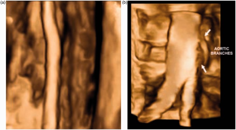
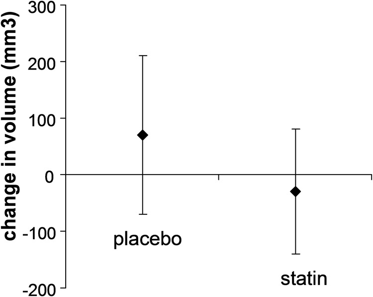
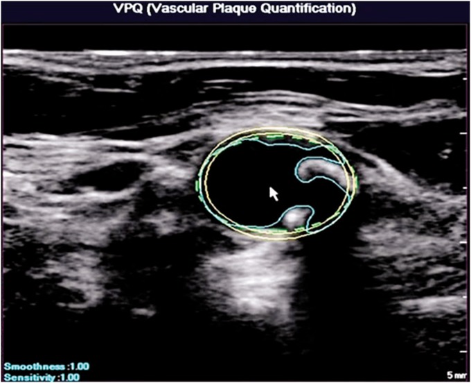
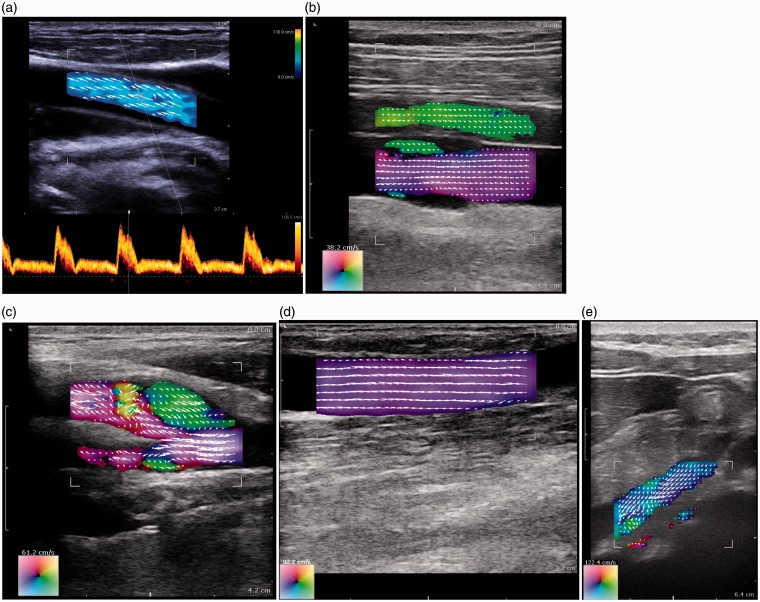
This article describes four technologies relevant to vascular ultrasound which are available commercially in 2015, and traces their origin back through the research literature. The technologies are 3D ultrasound and its use in plaque volume estimation (first described in 1994), colour vector Doppler for flow visualisation (1994), wall motion for estimation of arterial stiffness (1968), and shear wave elastography imaging of the arterial wall (2010). Overall these technologies have contributed to the understanding of vascular disease but have had little impact on clinical practice. The basic toolkit for vascular ultrasound has for the last 25 years been real-time B-mode, colour flow and spectral Doppler. What has changed over this time is improvement in image quality. Looking ahead it is noted that 2D array transducers and high frame rate imaging continue to spread through the commercial vascular ultrasound sector and both have the potential to impact on clinical practice.

Keywords: 3D; B-mode; Doppler ultrasound; elastography; plaque volume; shear wave imaging; spectral Doppler; stiffness; vascular ultrasound; vector Doppler; wall motion.


Figures

Figure 1
Figure 1
Surface-shaded views of (a) the femoral artery and (b) the aortic bifurcation. 3D ultrasound images are collected using the Philips Matrix array ultrasound scanner. Images provided courtesy of Royal Philips (Amsterdam, Netherlands)
Figure 2
Figure 2
Reduction in plaque volume after a course of statins; redrawn from data in Krasinski A, et al.
Figure 3
Figure 3
Automated segmentation of arterial wall and atherosclerotic plaque for use in 3D plaque volume estimation. Images provided courtesy of Royal Philips (Amsterdam, Netherlands)
Figure 4
Figure 4
Vector flow imaging using the BK 3000 scanner (BK Medical, Herlev, Denmark). (a) Common carotid artery with the automatic alignment of the angle cursor with the direction of blood motion. (b) Femoral artery and vein; notice the backflow behind the valve in the vein (bottom vessel). (c) Carotid artery with reverse flow in the bulb. (d) Well-functioning dialysis fistula. (e) Vena porta from a subcostal insonation window (Figure 4a provided by BK Medical. Figure 4b–e provided by Dr Kristoffer Hansen, Blegdamsvej, Denmark)
Figure 5
Figure 5
Increase in Young's modulus in carotid arteries in patients with peripheral arterial (PAD) compared to controls (redrawn from data in Claridge et al.
Figure 6
Figure 6
Wall motion obtained from TDI using a Philips HDI 5000 (Royal Philips, Amsterdam, Netherlands). (a) Healthy artery. Left top: TDI images in blue and red. Left middle: Instantaneous wall displacement as a function of longitudinal position along the artery. Left bottom: wall distension versus time. Right: Distension waveforms from all scan lines superimposed, plus mean distension (orange line). (b) TDI images in a diseased artery (red and blue); superimposed is the instantaneous wall distension as a function of longitudinal position in the artery
Figure 7
Figure 7
Wall motion obtained using an Esaote ultrasound system (Esaote, Genoa, Italy). Wall motion waveforms are shown at the bottom. The blue lines indicate the instantaneous wall distension as a function of longitudinal position in the artery (Courtesy Dr Peter Brands, Esaote)
Figure 8
Figure 8
Typical example of a stiffness image from a patient with carotid atherosclerosis. Reproduced from Ramnarine KV, Garrard JW, Kanber B, et al. Shear wave elastography imaging of carotid plaques: feasible, reproducible and of clinical potential. Cardiovascular Ultrasound 2014;12:49; published by BioMed Central.

References

    1. Fenster A, Parraga G, Bax J. Three-dimensional ultrasound scanning. Interface Focus 2011; 1: 503–19. - PMC - PubMed
    1. Prager RW, Ijaz UZ, Gee AH, et al. Three-dimensional ultrasound imaging. J Eng Med 2010; H2: 193–223. - PubMed
    1. Hammer S, Jeays A, MacGillivray TJ, et al. Acquisition of 3D arterial geometries and integration with computational fluid dynamics. Ultrasound Med Biol 2009; 35: 2069–83. - PubMed
    1. Smith SW, Trahey GE, von Ramm OT. Two-dimensional arrays for medical ultrasound. Ultrasonic Imaging 1992; 14: 213–33. - PubMed
    1. Light ED, Davidsen RE, Fiering JO, et al. Progress in two-dimensional arrays for real-time volumetric imaging. Ultrason Imag 1998; 20: 1–15. - PubMed