

Morphological and functional relationships with ultrasound measured muscle thickness of the lower extremity: a brief review
- PMID: 27433253
- PMCID: PMC4760590
- DOI: 10.1177/1742271X15587599
Morphological and functional relationships with ultrasound measured muscle thickness of the lower extremity: a brief review
Abstract
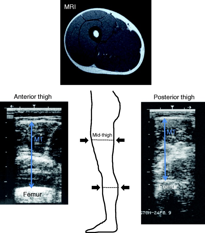
Ultrasound is a potential method for assessing muscle size of the extremity and trunk. In a large muscle, however, a single image from portable ultrasound measures only muscle thickness (MT), not anatomical muscle cross-sectional area (CSA) or muscle volume (MV). Thus, it is important to know whether MT is related to anatomical CSA and MV in an individual muscle of the extremity and trunk. In this review, we summarize previously published articles in the lower extremity demonstrating the relationships between ultrasound MT and muscle CSA or MV as measured by magnetic resonance imaging and computed tomography scans. The relationship between MT and isometric and isokinetic joint performance is also reviewed. A linear relationship is observed between MT and muscle CSA or MV in the quadriceps, adductor, tibialis anterior, and triceps surae muscles. Intrarater correlation coefficients range from 0.90 to 0.99, except for one study. It would appear that anterior upper-thigh MT, mid-thigh MT and posterior thigh MT are the best predictors for evaluating adductor, quadriceps, and hamstrings muscle size, respectively. Despite a limited number of studies, anterior as well as posterior lower leg MT appear to reflect muscle CSA and MV of the lower leg muscles. Based on previous studies, ultrasound measured anterior thigh MT may be a valuable predictor of knee extension strength. Nevertheless, more studies are needed to clarify the relationship between lower extremity function and MT.
Keywords: B-mode ultrasound; muscle CSA and volume; muscle thickness.
Figures
References
-
- Ikai M, Fukunaga T. Calculation of muscle strength per unit cross-sectional area of human muscle by means of ultrasonic measurement. Int Z Angew Physiol 1968; 26: 26–32. - PubMed
-
- Fukunaga T, Miyatani M, Tachi M, et al. Muscle volume is a major determinant of joint torque in humans. Acta Physiol Scand 2001; 172: 249–55. - PubMed
-
- Sanada K, Kearns CF, Kojima K, et al. Peak oxygen uptake during running and arm cranking normalized to total and regional skeletal muscle mass measured by magnetic resonance imaging. Eur J Appl Physiol 2005; 93: 687–93. - PubMed
-
- Narici MV, Roi GS, Landoni L. Force of knee extensor and flexor muscles and cross-sectional area determined by nuclear magnetic resonance imaging. Eur J Appl Physiol Occup Physiol 1988; 57: 39–44. - PubMed
Publication types
LinkOut - more resources
Full Text Sources
Medical